Chest spine ultrasonic scanning teaching method

A spine and thoracic technology, applied in the field of thoracic and spine ultrasound scanning teaching, can solve problems such as limited application, long learning curve for clinicians, and disordered scanning process

Inactive Publication Date: 2021-06-04
PEKING UNION MEDICAL COLLEGE HOSPITAL CHINESE ACAD OF MEDICAL SCI
View PDF0 Cites 0 Cited by
  • Summary
  • Abstract
  • Description
  • Claims
  • Application Information

AI Technical Summary

Problems solved by technology

This leads to disorder in the scanning process and inaccurate positioning of the target section for clinicians to learn about the ultrasound scan of the thoracic spine. In actual operation, it is not easy to understand the structure around the target section, ...

Method used


Image

  • Chest spine ultrasonic scanning teaching method
  • Chest spine ultrasonic scanning teaching method
  • Chest spine ultrasonic scanning teaching method

Examples


Embodiment Construction

[0020] In order to better understand the purpose, structure and function of the present invention, a teaching method for ultrasound scanning of the chest and spine of the present invention will be further described in detail below in conjunction with the accompanying drawings.

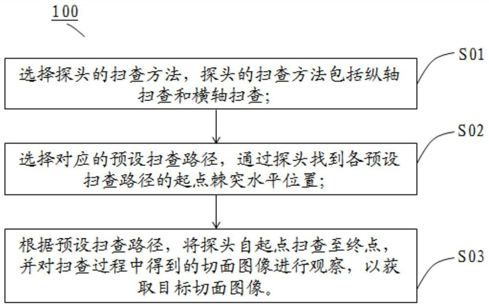
[0021] figure 1 A flow chart of the method 100 for teaching thoracic and spinal ultrasound scanning according to the present invention is shown. Such as figure 1 As shown, the teaching method 100 for thoracic and spinal ultrasound scanning includes: step one S01, select the scanning method of the probe, the scanning method of the probe includes vertical axis scanning and horizontal axis scanning; step two S02, select the corresponding The preset scanning path L, find the horizontal position of the spinous process at the starting point of each preset scanning path L through the probe; Step 3 S03, according to the preset scanning path L, scan the probe from the starting point to the end point, and scan ...


PUM


Abstract

The invention belongs to the field of ultrasonic scanning teaching methods, and discloses a chest spine ultrasonic scanning teaching method. The chest spine ultrasonic scanning teaching method comprises the following steps: step 1, selecting a scanning method of a probe, the scanning method of the probe comprising longitudinal axis scanning and transverse axis scanning; step 2, selecting corresponding preset scanning paths, and finding the starting point spinous process horizontal position of each preset scanning path through a probe; and step 3, according to a preset scanning path, scanning the probe from a starting point to an end point, and observing a section image obtained in the scanning process to obtain a target section image. The objective of the invention is to provide the teaching method for ultrasonic scanning of the chest spine, so that scanning guidance for clinicians is facilitated by establishing a scanning path diagram of the chest spine.

Description

technical field [0001] The invention belongs to the field of ultrasonic scanning teaching methods, in particular to a teaching method for thoracic and spine ultrasonic scanning. Background technique [0002] The spine of the human body includes cervical spine (usually represented by "C" in medicine), thoracic spine (thoracic spine, usually represented by "T" in medicine), lumbar spine (usually represented by "L" in medicine) and sacral vertebrae (usually represented by "L" in medicine). Denoted by "S"). Among them, there are 12 thoracic vertebrae, which are usually represented by "T" in medicine. From top to bottom, the vertebral body gradually increases. The transverse section of the vertebral body is heart-shaped, the upper thoracic vertebra is similar to the cervical vertebra, and the lower thoracic vertebra is similar to the lumbar vertebra. On the upper and lower margins of the posterior part of the vertebral body, there are upper and lower costal recesses that joint...

Claims


Application Information

Patent Timeline
IPC IPC(8): G09B23/28
CPCG09B23/286
Inventor 崔旭蕾费昱达谭刚张娇宋锴澄陈绍辉黄宇光
Owner PEKING UNION MEDICAL COLLEGE HOSPITAL CHINESE ACAD OF MEDICAL SCI
Why Eureka
Try Eureka