
Real-time pressure monitoring tracheal intubation device with suction function and method of real-time pressure monitoring tracheal intubation device

A technology for tracheal intubation and real-time monitoring, applied in the field of medical devices, can solve problems such as lack of drainage function, lung infection, and inability to monitor cuff pressure in real time, so as to avoid lung infection and be applicable to a wide range of scenarios.

Inactive Publication Date: 2022-03-04
ZHEJIANG UNIV
View PDF10 Cites 0 Cited by
  • Summary
  • Abstract
  • Description
  • Claims
  • Application Information

AI Technical Summary

Problems solved by technology

At present, in clinical practice, the pressure of the cuff is usually detected every 4 hours with a manometer, and the cuff leakage or even rupture cannot be detected in time.
In addition, in the current clinical practice, especially for patients who are bedridden for a long time in the intensive care unit and use a ventilator, it is easy for secretions such as saliva to flow into the trachea. When there is a gap between the cuff and the trachea due to other reasons, the secretion will penetrate into the trachea, which will easily lead to lung infection, increase the difficulty of treatment and the medical burden of patients and society
Facing the above major clinical needs, in view of the fact that the current endotracheal intubation device cannot monitor the cuff pressure in real time and has no drainage function, a solution for endotracheal intubation with real-time pressure monitoring and suction function is proposed

Method used


Image

  • Real-time pressure monitoring tracheal intubation device with suction function and method of real-time pressure monitoring tracheal intubation device
  • Real-time pressure monitoring tracheal intubation device with suction function and method of real-time pressure monitoring tracheal intubation device
  • Real-time pressure monitoring tracheal intubation device with suction function and method of real-time pressure monitoring tracheal intubation device

Examples


Embodiment Construction

[0030] The present invention will be further elaborated and illustrated below in conjunction with the accompanying drawings and specific embodiments. The technical features of the various implementations in the present invention can be combined accordingly on the premise that there is no conflict with each other.

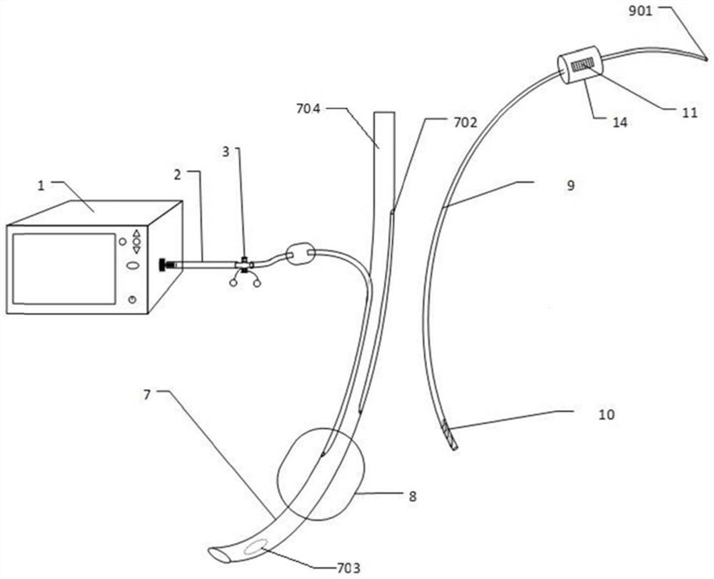
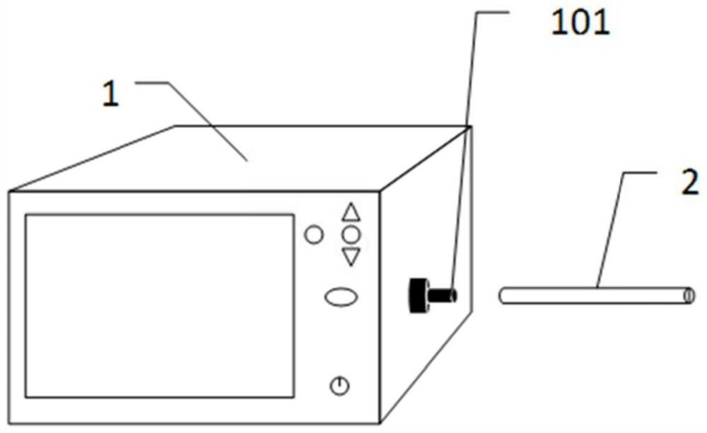
[0031] like figure 1 As shown, it is a real-time pressure monitoring endotracheal intubation device with suction function provided by the present invention. The endotracheal intubation device mainly includes an endotracheal tube 7 , a constant pressure inflation and deflation device 1 and an anti-blockage drainage tube 9 .

[0032] like Figure 4 As shown, the endotracheal tube 7 mainly includes a cannula 704 , an inflatable tube 701 and a cuff 8 . The intubation tube 704 is used to insert into the trachea of ​​the human body and assist the patient to breathe. The head of the intubation tube 704 is provided with a cuff 8, which can fix the intubation tube and sea...


PUM


Abstract

The invention discloses a real-time pressure monitoring trachea cannula device with a suction function and a method of the real-time pressure monitoring trachea cannula device. The trachea cannula device comprises an endotracheal catheter, a constant-pressure inflating and deflating device and an anti-blocking drainage tube. A first opening communicated with the air passage is formed in the side wall of the cannula located above the cuff, a second opening communicated with the outside is formed in the side wall of the cannula located outside the human body, and a drainage channel located in the cannula is connected between the first opening and the second opening; the anti-blocking drainage tube comprises a drainage end and a suction end, and the anti-blocking drainage tube can extend into the drainage channel through the second opening, so that the drainage end is arranged between the cannula and the airway through the first opening; the suction end is located outside the human body and can be connected with a suction device so that secretions in the air channel above the cuff can be sucked and emptied through the drainage end. Secretions in the airway above the cuff are sucked and emptied regularly through the drainage end, and pulmonary infection caused by the fact that the secretions in the airway penetrate into the trachea can be effectively avoided.

Description

technical field [0001] The invention belongs to the technical field of medical devices, and in particular relates to a real-time pressure monitoring endotracheal intubation device with a suction function and a method thereof. Background technique [0002] Endotracheal intubation is a method of inserting a special endotracheal tube through the oral cavity or nasal cavity and into the trachea or bronchus through the glottis to provide the best conditions for unobstructed airway, ventilation and oxygen supply, and airway suction. It is the first choice for rescuing patients with respiratory dysfunction. important measure. Endotracheal tubes generally consist of a tube and a cuff. Under normal circumstances, the pressure range of the cuff is generally 25-30mmHg. If it is too high, it will easily lead to hypoxia, ischemia and even necrosis of the tracheal wall mucosa. If it is too low, it will not be able to effectively close the gap between the cuff and the trachea. It is ther...

Claims


Application Information

Patent Timeline
Patent Type & Authority Applications(China)
IPC IPC(8): A61M16/04
CPCA61M16/044A61M16/0479A61M16/0486A61M2016/0027A61M2205/3331
Inventor 孙静卢如意何国军张倩冯靖祎蔡洪流
Owner ZHEJIANG UNIV
Learn More