Feature extraction method for frozen electron cross-sectional image

A technology of feature extraction and frozen electrons, which is applied in the field of structural biology, can solve the problems of affecting the accuracy of 3D reconstruction and reducing the number of particles, and achieve the effect of avoiding spatial movement errors

Pending Publication Date: 2022-04-15
ZHEJIANG UNIV
View PDF2 Cites 0 Cited by
  • Summary
  • Abstract
  • Description
  • Claims
  • Application Information

AI Technical Summary

Problems solved by technology

This method can improve the efficiency of particle selection, but this method needs to screen each image and remove some particles at the same time, so that the number of particles in the entire image set is reduced, which affects the accuracy of subsequent 3D reconstruction

Method used


Image

  • Feature extraction method for frozen electron cross-sectional image
  • Feature extraction method for frozen electron cross-sectional image
  • Feature extraction method for frozen electron cross-sectional image

Examples


Embodiment Construction

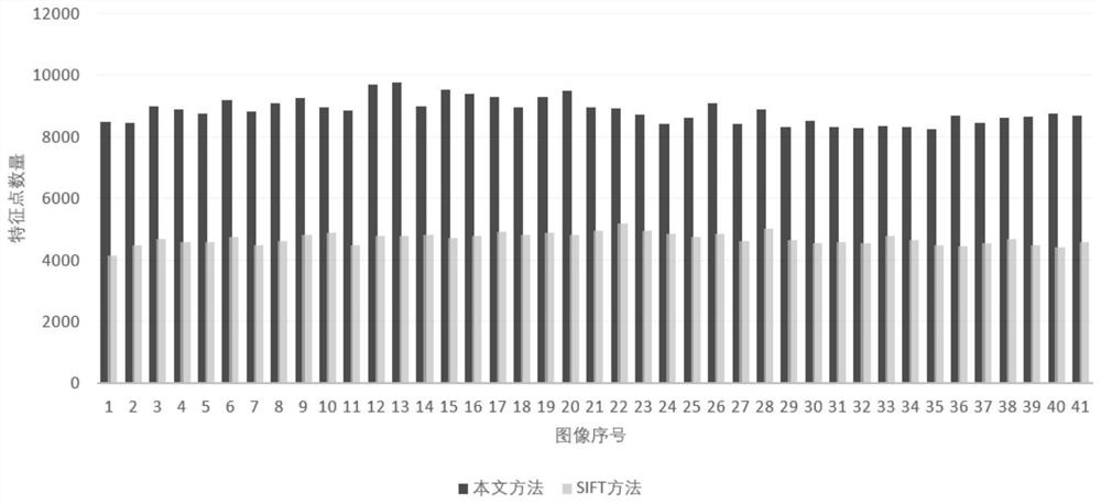
[0032] A set of experimental data is set, the projection angle ranges from -60° to 60°, and the interval of each projection is 3°. A total of 41 projection images of the original cryo-electron tomography are taken, each with a size of 5760×4092.

[0033] For this set of data, such as figure 1 As shown, the feature extraction of the cryo-electron tomography image is performed, including:

[0034] S1 acquires the above experimental data, including its projection angle range, shooting interval and corresponding image sequence;

[0035] S2 is based on a projection angle range of -60° to 60°, a projection interval of 3°, and a preset angle sequence (0,3°,6°,…60°).

[0036] S3 performs plane affine transformation on each cryo-electron tomogram in the image sequence based on the preset angle sequence (0, 3°, 6°,...60°):

[0037] Calculate the tilt parameter first:

[0038]

[0039] Where t is the tilt parameter and θ is the tilt angle.

[0040] Based on the above calculation r...


PUM


Abstract

The invention discloses a feature extraction method for a frozen electron cross-sectional image. The method comprises the following steps: S1, acquiring a projected image of a frozen electron cross-sectional image and a corresponding image sequence; s2, presetting an angle sequence of affine transformation simulation; s3, according to a set angle sequence, affine transformation simulation is carried out on the projection image of each frozen electron tomography, and a corresponding simulation image set is obtained after screening; and S4, according to the analog image set obtained in the S3 and the corresponding angle sequence, extracting and obtaining matched feature points. According to the method, through the simulated image set and the corresponding angle sequence, the interference on the spatial position of the sample during manual tilting is solved, and the extraction quality of the feature points is improved, so that a high-quality frozen electron three-dimensional reconstruction model is obtained.

Description

technical field [0001] The invention relates to the technical field of structural biology, in particular to a feature extraction method for frozen electron tomography images. Background technique [0002] Electron tomography three-dimensional reconstruction technology is an important branch of using cryo-electron microscopy to study the structure of biological macromolecules. For the study of amorphous, asymmetric and non-uniform biological sample structures, it has irreplaceable advantages of other cryo-electron microscopy reconstruction technologies. [0003] The traditional electron tomography three-dimensional reconstruction technology reversely reconstructs the three-dimensional structure of the same biological sample by obtaining two-dimensional projection images under multiple continuous angles. During the projection imaging process, the number of electrons emitted by the transmission electron microscope is The direction is fixed, and the sample is tilted along an axi...

Claims


Application Information

Patent Timeline
IPC IPC(8): G06V10/46G06V10/75G06K9/62
Inventor 冯结青郭珂诚
Owner ZHEJIANG UNIV
Why Eureka
Try Eureka