Reset fixing device and method for distal tibiofibular syndesmosis separation

A fixation device, tibiofibular technology, applied in the field of medical devices, can solve the problems of non-conformity with biomechanics, poor separation and reduction of the lower tibiofibular syndesmosis, displacement of lateral malleolus fractures, etc., so as to improve the reduction and fixation effect and meet the requirements of biomechanics Effect

Active Publication Date: 2022-05-13
TONGDE HOSPITAL OF ZHEJIANG PROVINCE
View PDF10 Cites 0 Cited by
  • Summary
  • Abstract
  • Description
  • Claims
  • Application Information

AI Technical Summary

Problems solved by technology

The traditional treatment method uses point-type reduction forceps to clamp the inferior tibiofibular syndesmosis for reduction, and then drive one or two fixation screws from the lateral malleolus to the distal end of the tibia. However, in the process of reducing the tibiofibular syndesmosis separation, due to the uneven force on the lateral malleolus, poor reduction of the inferior tibiofibular syndesmosis or even displacement of the lateral malleolus fracture may occur, and the traditional point-type reduction forceps are prone to forceps arms during the reduction process. The skin is pinched, especially for thick legs, and the length of the needle insertion point from the ankle joint plane is roughly estimated. If the position of the fixation screw does not meet the biomechanical requirements, the fixation effect is not good or even the nail breaks.

Method used


Image

  • Reset fixing device and method for distal tibiofibular syndesmosis separation
  • Reset fixing device and method for distal tibiofibular syndesmosis separation
  • Reset fixing device and method for distal tibiofibular syndesmosis separation

Examples


Embodiment

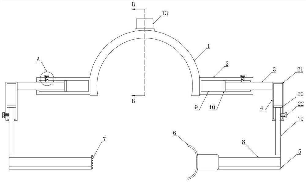
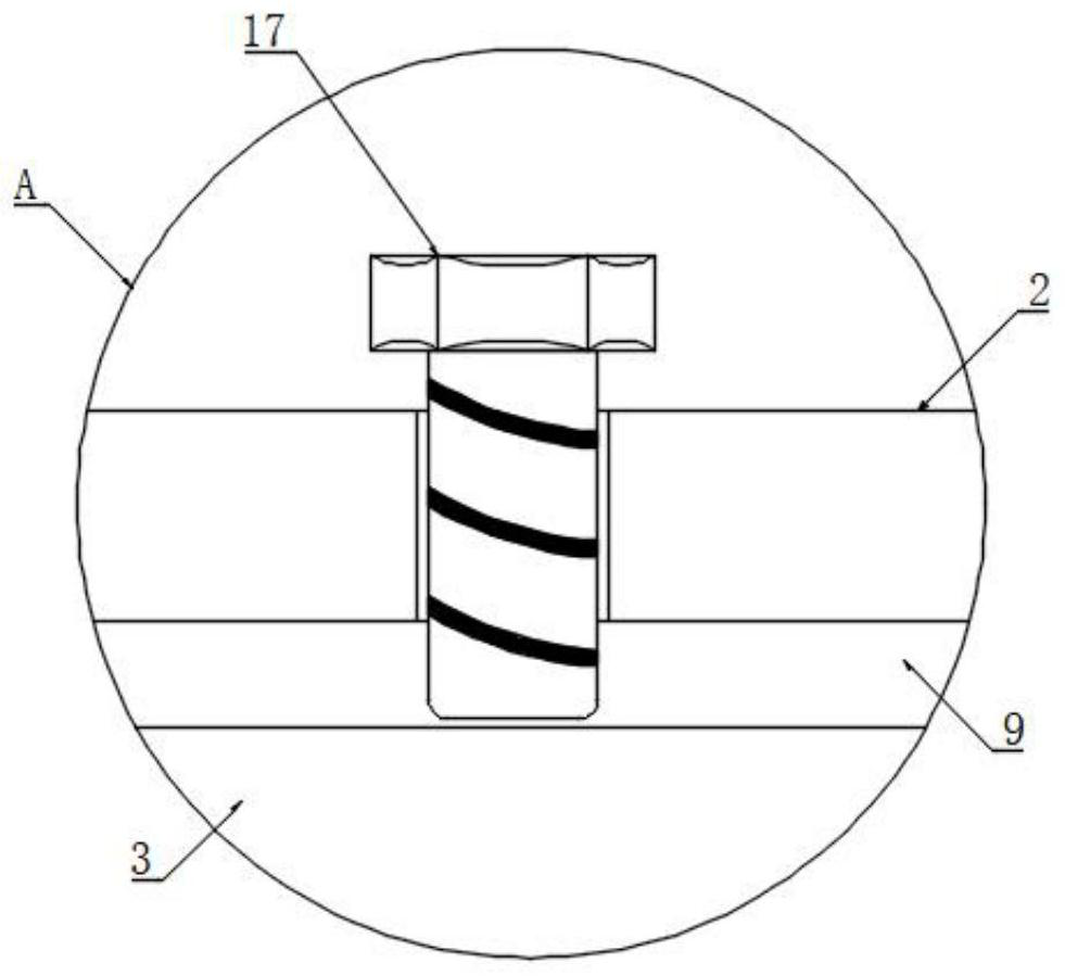
[0024] Embodiment: a kind of reduction and fixation device for the separation of inferior tibiofibular syndesmosis, as attached figure 1 And attached figure 2 As shown, the angle plate 1 is included, and the angle plate 1 is in an arc-shaped structure. The first fixed rod 2 is arranged on both sides of the angle plate 1. The first fixed rod 2 is provided with a first cavity 9. The first cavity 9 A first moving rod 3 is provided inside, and a first fixing bolt 17 is arranged on the first fixing rod 2, and the first fixing bolt 17 extends into the first cavity 9, and the first fixing bolt 17 is set for fixing the first The position of the moving rod 3 is fixed, and one end of the first moving rod 3 located in the first cavity 9 is provided with a first limiting plate 10, and the first limiting plate 10 is slidably matched with the first cavity 9 to set the first limiting plate 10. A limit plate 10 can prevent the first moving rod 3 from being completely withdrawn from the firs...


PUM


Abstract

The invention discloses a reduction fixing device and method for distal tibiofibular syndesmosis separation.The device comprises an angle scale, first fixing rods are symmetrically arranged on the two sides of the angle scale, first cavities are formed in the first fixing rods, first moving rods are arranged in the first cavities, and the first moving rods extend out of the first cavities and are connected with first vertical rods; a third cavity is formed in the first vertical rod, a first sliding rod is arranged in the third cavity, the first sliding rod extends out of the third cavity and is connected with a hollow guider, and an inner cavity is formed in the hollow guider; the method comprises the following steps of longitudinal pre-positioning, transverse pre-positioning and transverse fixing. According to the invention, accurate reduction and fixation in different dimensions can be carried out on inferior tibiofibular syndesmosis separation, the requirements of biomechanics are met, and reduction and fixation can be carried out on lateral malleolus fracture at the same time.

Description

technical field [0001] The invention relates to the technical field of medical devices, in particular to a reduction and fixation device and method for the separation of inferior tibiofibular syndesmosis. Background technique [0002] Separation of the inferior tibiofibular syndesmosis is more common in clinical practice, often accompanied by fractures of the lateral malleolus, and how well it is handled directly affects the function of the ankle joint. The traditional treatment method uses point-type reduction forceps to clamp the inferior tibiofibular syndesmosis for reduction, and then drives one or two fixation screws from the lateral malleolus to the distal end of the tibia. However, in the process of reducing the tibiofibular syndesmosis separation, due to the uneven force on the lateral malleolus, poor reduction of the inferior tibiofibular syndesmosis or even displacement of the lateral malleolus fracture may occur, and the traditional point-type reduction forceps ar...

Claims


Application Information

Patent Timeline
Patent Type & Authority Applications(China)
IPC IPC(8): A61B17/66
CPCA61B17/66Y02A50/30
Inventor 朱佳福
Owner TONGDE HOSPITAL OF ZHEJIANG PROVINCE
Why Eureka
Try Eureka