PD-L1-targeting polypeptide PET molecular imaging probe as well as preparation method and application thereof

A technology of PD-L1 and molecular imaging, applied in the fields of radiopharmaceutical chemistry and clinical nuclear medicine, to achieve rapid metabolism and excellent in vivo targeting performance

Pending Publication Date: 2022-06-03
HARBIN MEDICAL UNIVERSITY
View PDF4 Cites 2 Cited by
  • Summary
  • Abstract
  • Description
  • Claims
  • Application Information

AI Technical Summary

Problems solved by technology

As a result, it takes many days to clear molecular imaging probes from no

Method used


Image

  • PD-L1-targeting polypeptide PET molecular imaging probe as well as preparation method and application thereof
  • PD-L1-targeting polypeptide PET molecular imaging probe as well as preparation method and application thereof
  • PD-L1-targeting polypeptide PET molecular imaging probe as well as preparation method and application thereof

Examples


Embodiment 1

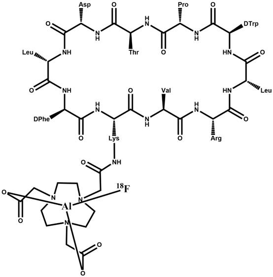
1) Preparation of NOTA-PA2881: 1. Dissolve 5 mg Cyclopeptide 66 in HEPES-EDTA solution, add 3 equivalents of triisopropylacetamide (2-carboxyethyl) phosphine (dissolved in 0.2 mol / L) ammonium acetate buffer, pH=7), evacuated the gas in the solution, repeated three times; reacted at room temperature for 60 minutes, then transferred to a centrifugal filter, centrifuged at 4000 rpm for 90 minutes; 1 mL of 0.2 mol / L ammonium acetate buffer The reduced peptide was added to 2 mL of 0.2 mol / L ammonium acetate buffer (pH=7) and transferred to the second reaction vessel; 4 mg of NOTA-maleimide and 0.5 mL of 0.2 mol / L The sodium acetate buffer (pH=7) was mixed and added to the second reaction vessel, and reacted at 40 °C for 3 hours; after the reaction was completed, the reaction mixture was transferred to a centrifugal filter, and centrifuged at 4000 rpm for 90 minutes; after discarding the flow , add 2 mL of ultrapure water, centrifuge again at 4000 r / min for 90 minutes, and discard th...

Embodiment 2

probe 18 In vivo targeting validation of F-AlF-NOTA-PA2881.

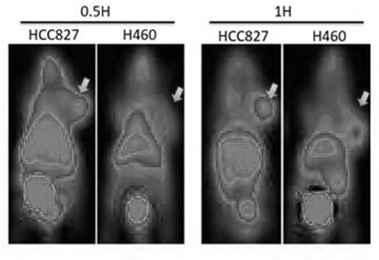
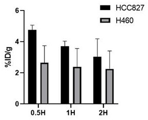
[0037] In the in vivo targeting validation experiment, the cell line HCC827 with high PD-L1 expression and the cell line H460 with low PD-L1 expression were used to establish a nude mouse tumor model (female mouse, 5 weeks old, about 20 g, 2 × 10 6 The number of cells was subcutaneously implanted on the right shoulder, and the tumor volume was about 300mm after about 4 weeks 3 in vivo imaging experiments), injected via mouse tail 18 After F-AlF-NOTA-PA2881 probe solution (injection dose is 9.25MBq, 150 μL), 0.5 hour, 1 hour PET / CT scan (Discovery 790Elite, GE healthcare) was performed under isoflurane-oxygen anesthesia. The duration is 5 minutes, and the image post-processing platform AW4.6 software (GE Healthcare).

[0038] Imaging results such as figure 1 , HCC827 tumor uptake probe was significantly higher than background and significantly higher than H460, indicating that the probe 18 F-AlF-NOTA-PA2881 can s...


PUM


Abstract

The invention relates to the technical field of radiopharmaceutical chemistry and clinical nuclear medicine, in particular to a PD-L1 targeting polypeptide PET molecular imaging probe and a preparation method and application thereof. The < 18 > F-AlF-NOTA-PA2881 probe is an annular polypeptide PET (Polyethylene Terephthalate) molecular imaging probe composed of ten amino acids based on Cyclopetide 66, and is formed by connecting a bifunctional chelating agent to lysine in cyclic peptide and then labeling with radionuclide < 18 > F. The invention also discloses a preparation method of the < 18 > F-AlF-NOTA-PA2881 probe. And by adopting the short-half-life radionuclide 18F marker, the tumor focus can be quickly and accurately identified. Compared with the existing mainstream monoclonal antibody imaging probe, the molecular imaging probe has the advantages that the metabolism is rapid, and the potential influence of radionuclides and monoclonal antibodies on organisms is further reduced. By means of in-vitro imaging equipment PET, the expression level of tumor cells PD-L1 can be reflected, the immunotherapy curative effect aiming at the signal channel can be predicted, real-time curative effect detection, prognosis evaluation and the like can be achieved. The in-vivo targeting performance of the probe is excellent.

Description

technical field [0001] The invention relates to the technical fields of radiopharmaceutical chemistry and clinical nuclear medicine, namely a polypeptide PET molecular imaging probe targeting PD-L1 and a preparation method and application thereof. Background technique [0002] In the prior art, immune checkpoints are signaling pathways that play an important role in mediating immune tolerance and preventing excessive immune responses, among which programmed death receptor-1 (PD-1) / programmed death ligands 1 and 2 (PD-L1 / 2) signaling pathway can be expressed in a variety of malignant tumor tissues, affecting antigen signal presentation. PD-1 is a negative costimulatory receptor expressed on the surface of activated T cells and primary B cells. PD-1 has two ligands (PD-L1 / 2). PD-L1 is a widely expressed T cell, A type 1 transmembrane surface glycoprotein ligand in B cells and macrophages, PD-1 interacts with PD-L1 and negatively regulates immunity by regulating T lymphocyte p...

Claims


Application Information

Patent Timeline
IPC IPC(8): A61K51/08A61K51/04C07K7/64C07K1/13A61K101/02
CPCC07K7/64A61K51/0482A61K51/08
Inventor 孙夕林王竞胡欣欣韩兆国王凯杨丽丽刘想孙颖李晓娜王洪斌
Owner HARBIN MEDICAL UNIVERSITY
Why Eureka
Try Eureka