Cell nucleus segmentation method based on region enhancement
A technology of fine segmentation of cell nuclei, applied in image enhancement, neural learning methods, image analysis, etc., to achieve the effect of improving accuracy
Pending Publication Date: 2022-06-10
深圳市东汇精密机电有限公司
View PDF0 Cites 2 Cited by
- Summary
- Abstract
- Description
- Claims
- Application Information
AI Technical Summary
Problems solved by technology
[0004] During the implementation of this application, the inventors found that there are at least the following problems in this technology: the traditional cell nucleus segmentation method relies on the difference between the nucleus and the background or depends on the difference between the nucleus and the background. Predefine geometric shapes to generate markers; relying on the difference between nuclei and background will produce unreliable results and is not suitable for images of challenging scenarios such as nuclei crowding, overlapping, occlusion, etc., making it difficult to accurately segment the boundaries of nuclei; Relying on the predefined geometry of nuclei to generate markers can lead to less accurate segmentation due to large differences in shape, size, and chromatin pattern in nuclei of different cell types or different disease types Difficulty at the border of the cell nucleus
Method used
View moreImage
Smart ImageViewing Examples
Examples
Embodiment Construction
Login to View More PUM
 Abstract
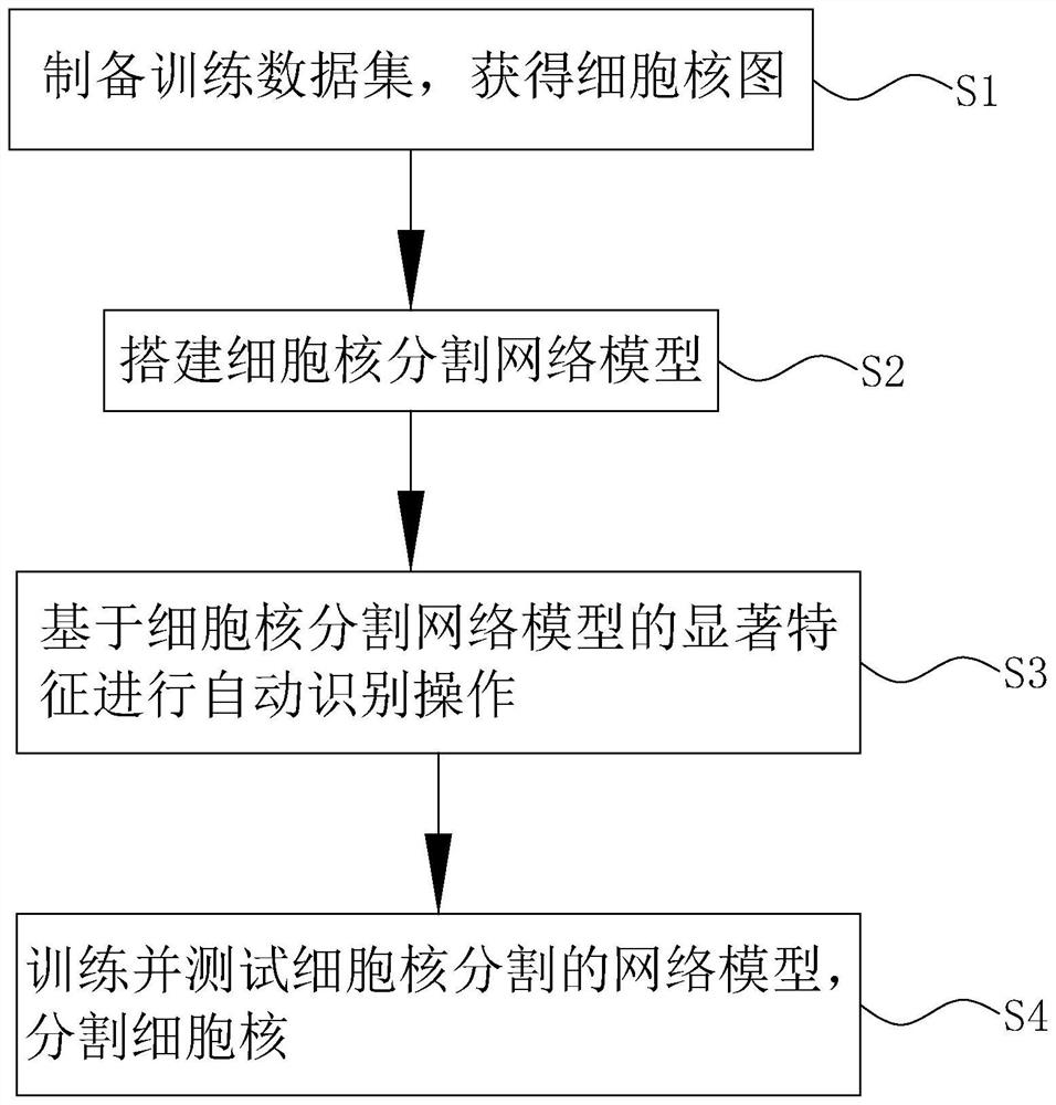
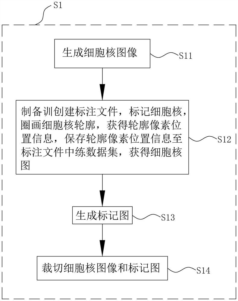
The invention discloses a cell nucleus segmentation method based on region enhancement, and belongs to the field of intelligent pathological diagnosis, and the method comprises the steps: preparing a training data set, and obtaining a cell nucleus image; the method comprises the following steps: constructing a cell nucleus segmentation network model, loading a cell nucleus image, and obtaining highlighted significant features, the cell nucleus segmentation network model comprising a contour branch used for detecting the edge of a cell nucleus and generating contour features, and a coarse segmentation branch used for completing a semantic segmentation task of the nucleus and predicting a cell nucleus proposal area in combination with the contour branch; the fine segmentation branch is used for processing the picture with the enhanced area; performing automatic identification operation based on the significant features of the cell nucleus segmentation network model; and training and testing the network model of cell nucleus segmentation, and segmenting the cell nucleus. The boundary of the cell nucleus can be accurately segmented.
Description
technical field [0001] The invention relates to the technical field of intelligent pathological diagnosis, in particular to a cell nucleus segmentation method based on region enhancement. Background technique [0002] Cervical cancer is posing a huge threat to the health of women all over the world, and many people in the world die because of cervical cancer. To study cervical cancer, cytopathologists need to screen for abnormal cells, but screening for abnormal cells from cervical cytology specimens is a tedious and laborious task, therefore, it is necessary to develop automated screening techniques to assist cytopathologists Diagnose the cervical smear. Among them, nuclei segmentation is a key task for automated screening and diagnosis, as nuclei provide chromatin richness and nuclei morphological information. [0003] Image segmentation technology based on deep learning is widely used in various fields of medical images, such as CT, MRI, pathological images, etc.; in th...
Claims
Login to View More Application Information
Patent Timeline
 Patent Type & Authority Applications(China)
IPC IPC(8): G06T7/11G06N3/04G06N3/08
CPCG06T7/11G06N3/08G06T2207/20081G06T2207/30024G06T2207/30204G06N3/048
Inventor 何彩梅刘锦烽罗志清陈建华蔡顺华刘钰凡何勇军
Owner 深圳市东汇精密机电有限公司
Features
- R&D
- Intellectual Property
- Life Sciences
- Materials
- Tech Scout
- Unparalleled Data Quality
- Higher Quality Content
- 60% Fewer Hallucinations
Learn More Browse by: Latest US Patents, China's latest patents, Technical Efficacy Thesaurus, Application Domain, Technology Topic, Popular Technical Reports.
© 2025 PatSnap. All rights reserved.Legal|Privacy policy|Modern Slavery Act Transparency Statement|Sitemap|About US| Contact US: help@patsnap.com


