
Method and control device to operate a magnetic resonance tomography apparatus

A technology of magnetic resonance tomography and control devices, which is applied in the direction of radiological diagnostic instruments, magnetic resonance measurement, and measurement using nuclear magnetic resonance image systems, which can solve problems such as matching and achieve the effect of improving quality

Inactive Publication Date: 2005-08-17
SIEMENS AG
View PDF5 Cites 12 Cited by
  • Summary
  • Abstract
  • Description
  • Claims
  • Application Information

AI Technical Summary

Problems solved by technology

However, the problem with all these methods is how to match each model with the geometric information obtained from the photo of the rough image

Method used


Image

  • Method and control device to operate a magnetic resonance tomography apparatus
  • Method and control device to operate a magnetic resonance tomography apparatus
  • Method and control device to operate a magnetic resonance tomography apparatus

Examples


Embodiment Construction

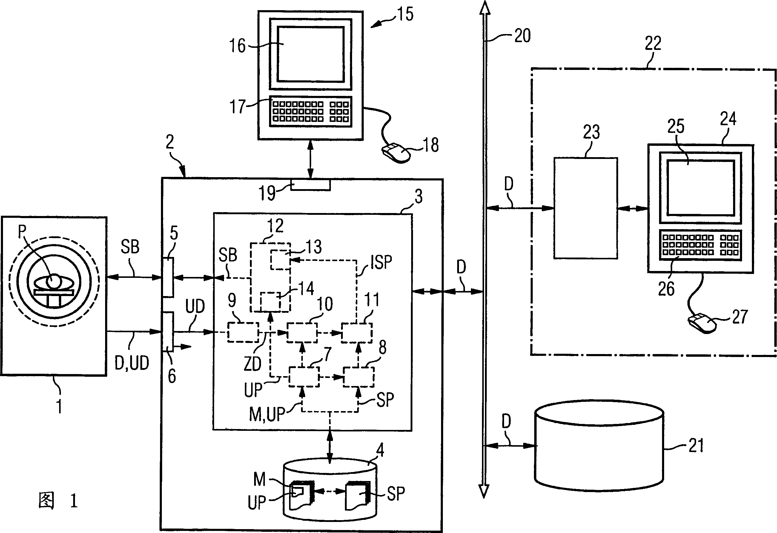
[0049] FIG. 1 shows an embodiment of a magnetic resonance tomography system 1 according to the invention with an associated control device 2 according to the invention connected to a bus 20 . Other components such as a mass memory 21 for storing image data D and a workstation 22 are connected to the bus 20 . The workstation 22 consists of a graphics computer 23 and a console 24 which typically has a display screen 25 as a user interface, a keyboard 26 and a pointing device 27 (such as a mouse). Workstation 22 is used, for example, for subsequent viewing and processing of the images produced by MRT system 1 .

[0050] Naturally, in the case of forming a larger network, existing components in other general radiology information systems (RIS), such as other functional blocks, mass memory, workstations, output devices (such as printers) can also be connected to the bus 20 , film production stations, and more. Also, it is possible to connect with external network or other RIS. I...


PUM


Abstract

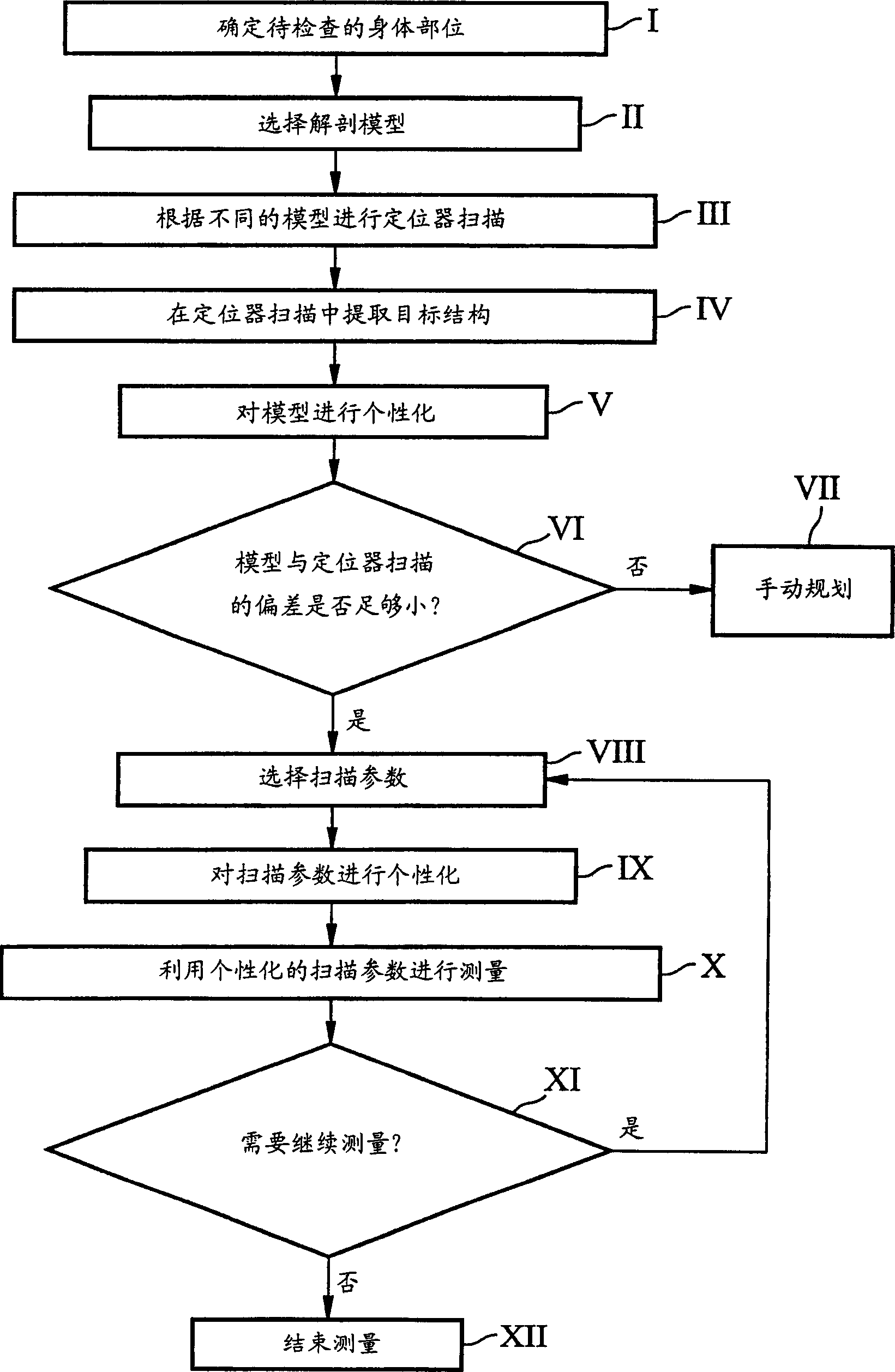
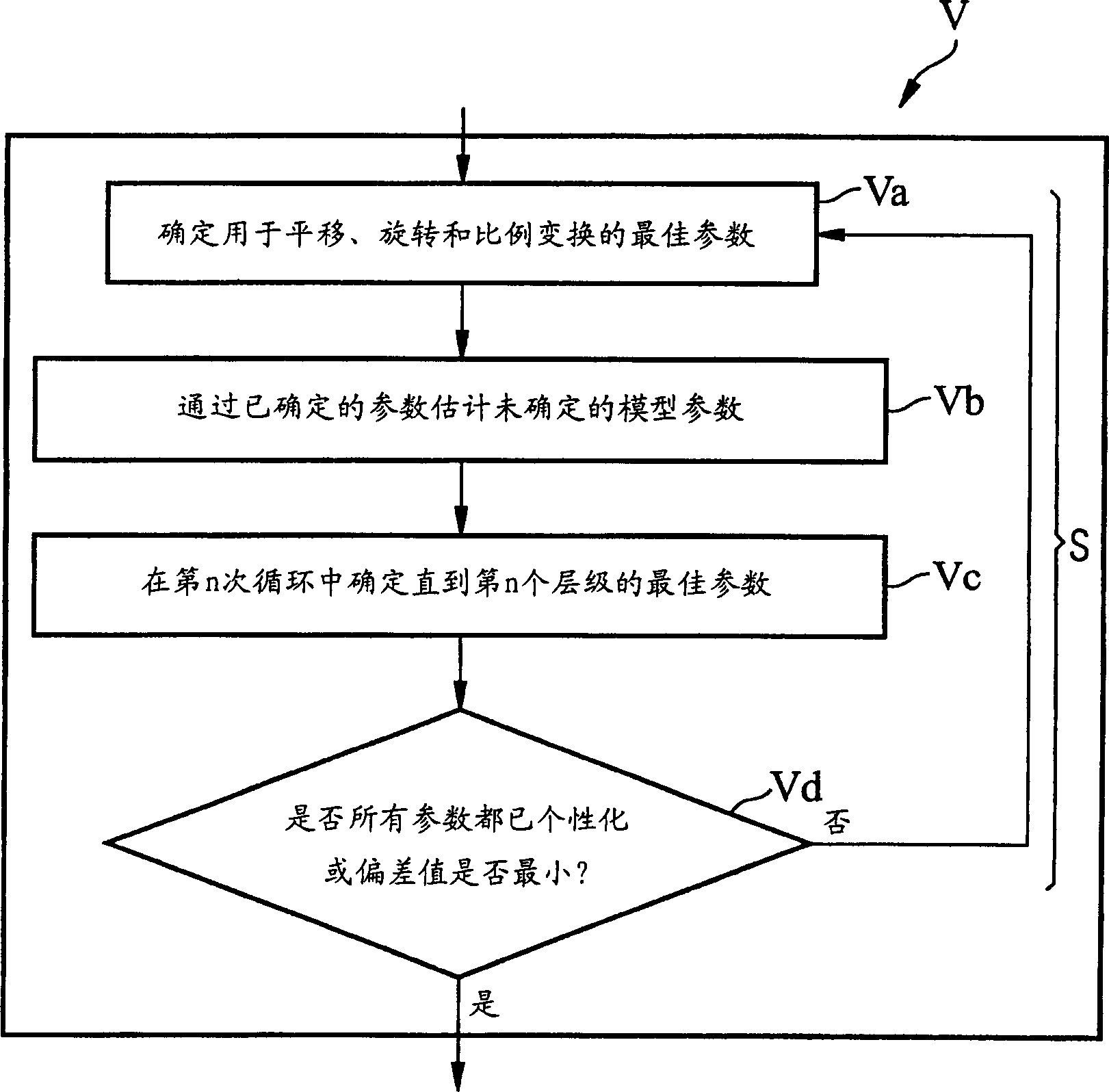
The present invention relates to a method for operation of resonance dislocation image making equipment.Firstly, a dissection canonical model (M) with changeable geometric figure is selected for an examination object depending on diagnosis problems.And then, a plurality of compendia images are measured for areas including the examination object, and different compendia sweep parameters (UP) used for controlling measurement of the compendia images are determined according to the chosen dissection canonical model (M).An object construction (Z) is determined in the sectional view image data (UD) of the measured compendia images, and the canonical model (M) is personalized to match the object construction (Z).According to the chosen canonical model (M) and the diagnosis problems, subsequent dislocation images sweep parameters (SP) for the resonance equipment are selected and controlled, the chosen sweep parameters (SP) are personalized corresponding to the personalized canonical model (M), and a plurality of dislocation images are measured on the basis of the personalized sweep parameter (ISP). The invention also relates to a corresponding image processing system.

Description

technical field [0001] The invention relates to a method for operating a magnetic resonance tomography system (MRT system). Furthermore, the invention relates to a corresponding control device for operating a magnetic resonance tomography system. Background technique [0002] The results of magnetic resonance tomography examinations are usually sequences with sections of the object of interest (e.g. the patient's head, knees, pelvis or a specific organ) or stretched areas of the patient belonging to the object of interest image (tomographic image). Here, the planning of the examination, i.e. the determination of the different "scanning parameters", e.g. position and number of slice stacks or individual slices, distance of slices to each other, volume, observation window and size of the measurement matrix or saturation area, etc., This is usually achieved through the interaction of the plant operator. Usually, when starting the measurement, first a broad overview image of ...

Claims


Application Information

Patent Timeline
Patent Type & Authority Applications(China)
IPC IPC(8): A61B5/055G01R33/54
CPCA61B6/488A61B5/055G01R33/546A61B6/545G01R33/54
Inventor 马丁·坦克
Owner SIEMENS AG
Learn More