Combined ultrasonic imaging and spectroscopic molecular analysis

A spectroscopic analysis, ultrasound imaging system technology, applied in the field of combined ultrasound imaging and spectroscopic molecular analysis, capable of solving problems such as the impossibility of specifying the properties of blood in detail

Inactive Publication Date: 2007-05-23
KONINKLIJKE PHILIPS ELECTRONICS NV
View PDF1 Cites 6 Cited by
  • Summary
  • Abstract
  • Description
  • Claims
  • Application Information

AI Technical Summary

Problems solved by technology

However, with ultrasound imaging it is often not possible to detail certain properties of the blood, such as th

Method used


Image

  • Combined ultrasonic imaging and spectroscopic molecular analysis
  • Combined ultrasonic imaging and spectroscopic molecular analysis
  • Combined ultrasonic imaging and spectroscopic molecular analysis

Examples


Embodiment Construction

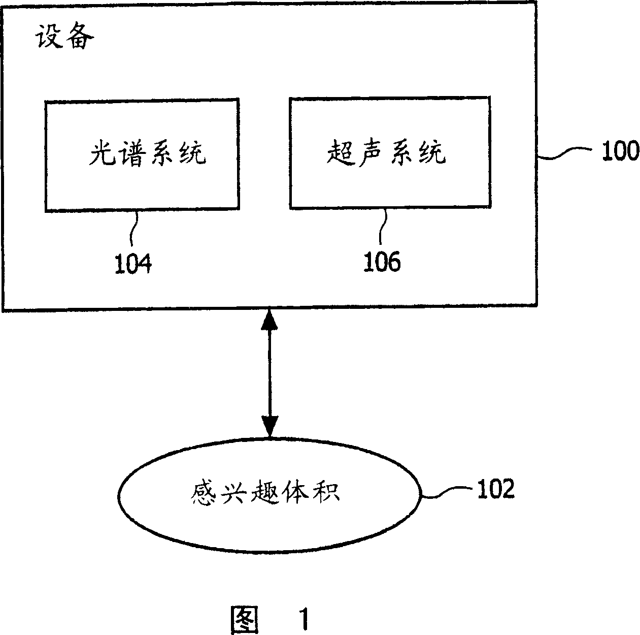
[0041] FIG. 1 illustrates a block diagram of an apparatus 100 including a spectroscopy system 104 and an ultrasound system 106 . The device 100 is adapted to investigate a volume of interest 102 . The ultrasound system 106 of the device 100 is adapted to acquire ultrasound echo signals from the volume of interest to generate a visual representation of the volume of interest 102 . In turn, the spectroscopic system 104 is adapted to perform spectroscopic analysis of biological structures located within the volume of interest 102 . Spectroscopy system 104 is preferably suitable for, but not limited to, Raman spectroscopy, for example. Other optical spectroscopy techniques can also be used. These include various methods based on Raman scattering, including nonlinear Raman spectroscopy, such as stimulated Raman spectroscopy and coherent anti-Stokes Raman spectroscopy, infrared spectroscopy, especially infrared absorption spectroscopy, Fourier Transform infrared spectroscopy and ...


PUM


Abstract

The invention provides an apparatus for combined imaging and analyzing of a biological structure by means of ultrasonic and spectroscopic techniques. The inventive diagnostic apparatus is adapted for simultaneous acquisition of ultrasonic echo signals and surface enhanced Raman spectroscopic signals. The invention makes particular use of a contrast agent that on the one hand provides enhanced reflection efficiency for ultrasonic echo signals and on the other hand enables surface enhanced Raman spectroscopy. The contrast agent comprises conventional microbubbles as well as solid metal nano-particles or combinations thereof. The invention therefore effectively provides non-invasive in vivo analysis of blood as well as detection and visualization of a blood flow.

Description

technical field [0001] The present invention relates to the field of imaging and analyzing biological structures using ultrasound and spectroscopy techniques. Background technique [0002] Within the framework of medical diagnosis, blood analysis is of great importance. Blood analysis involves information on blood flow within the cardiovascular system and the analysis of a patient's blood composition. To detect and visualize the flow of blood, sonography and ultrasound imaging are now successfully utilized. These ultrasound imaging techniques effectively utilize an ultrasound contrast agent (UCA) injected into the patient's bloodstream. Typically, UCAs are based on gas-filled microbubbles with pronounced reflective properties to ultrasound. Thus, a patient's blood flow can be effectively detected and visualized by injecting a contrast agent into the patient's cardiovascular system and subsequently using an ultrasound imaging system. Blood flow information is very importa...

Claims


Application Information

Patent Timeline
IPC IPC(8): G01N29/06G01N21/65A61B8/12G01N21/62A61B8/06G01J3/44A61B5/00G01N33/48A61B5/103G01S15/89A61B5/145
CPCG01N2291/02836G01N2291/02475G01N2291/02483G01N2291/044G01N29/06G01N2291/02466G01N2291/02433
Inventor G·卢卡森N·威拉尔德S·尼尔肯T·范邦默尔
Owner KONINKLIJKE PHILIPS ELECTRONICS NV
Why Eureka
Try Eureka