Electrocautery hemostasis clip

a hemostasis and uterus technology, applied in the field of electrocautery hemostasis clips, can solve the problems of increasing the risk of perforation or damage to the wall of the gi tract, and conventional tissue closure devices may not be sufficient to close certain tissue defects

Active Publication Date: 2019-06-04
BOSTON SCI SCIMED INC
View PDF13 Cites 607 Cited by
  • Summary
  • Abstract
  • Description
  • Claims
  • Application Information

AI Technical Summary

Problems solved by technology

These procedures may increase the risk of perforating or damaging the wall of the GI tract, or may require closure of the GI tract wall as part of the procedure.
However, conventional tissue closure devices may be insufficient to close certain tissue defects.

Method used


Image

  • Electrocautery hemostasis clip
  • Electrocautery hemostasis clip
  • Electrocautery hemostasis clip

Examples


Embodiment Construction

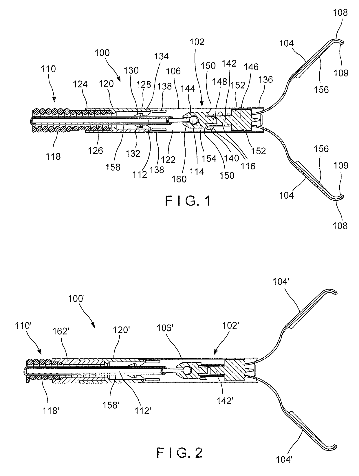
[0029]The present disclosure may be further understood with reference to the following description and the appended drawings, wherein like elements are referred to with the same reference numerals. The present disclosure is directed to an endoscopic clipping device for treating tissue perforations, defects and / or bleeds. In particular, exemplary embodiments of the present disclosure describe a hemostatic clip having both clipping and coagulation capabilities. Portions of the clipping device may be insulated or formed of non-conductive material to achieve a desired coagulating effect. It should be noted that the terms “proximal” and “distal,” as used herein, are intended to refer to a direction toward (proximal) and a direction away from (distal) a user of the device.

[0030]As shown in FIG. 1, a device 100 according to an exemplary embodiment of the present disclosure comprises a distal portion 102 insertable into a living body through, for example, a working channel of an endoscope t...


PUM

PropertyMeasurementUnit
thicknessaaaaaaaaaa
thicknessaaaaaaaaaa
thicknessaaaaaaaaaa

Abstract

A device for treating a tissue includes a capsule extending longitudinally from a proximal end to a distal end and including a channel extending therethrough, the capsule releasably coupled to a proximal portion of the device and clip arms, proximal ends of which are slidably received within the channel of the capsule so that the clip arms are movable between an open configuration, and a closed configuration. A core member is coupled to the clip arms, the core member including a proximal portion and a distal portion releasably connected to one another so that, when the core member is subjected to a predetermined load, the proximal and distal portions are separated from one another. An electrically conductive control member is connected to the core member, a proximal end of the connected member connected to a power source for delivering an electrical current to the clip arms.

Description

PRIORITY CLAIM[0001]This application claims priority to U.S. Provisional Patent Application Ser. No. 62 / 262,729 filed Dec. 3, 2015; the disclosure of which is incorporated herewith by reference.BACKGROUND[0002]Physicians have become more willing to perform more aggressive interventional and therapeutic endoscopic procedures including, for example, removal of larger lesions (e.g., cancerous masses), tunneling under mucosal layers in the gastro-intestinal (GI) tract to treat tissues below the mucosa, full thickness removal of tissue, inserting devices through the GI tract and then penetrating the GI organ to treat tissue outside the GI tract, and endoscopic treatment / repair of post-surgical issues (e.g., post-surgical leaks, breakdown of surgical staple lines, anastomotic leaks). These procedures may increase the risk of perforating or damaging the wall of the GI tract, or may require closure of the GI tract wall as part of the procedure. Endoscopic closure reduces cost and may reduce...

Claims


Application Information

Patent Timeline
Patent Type & Authority Patents(United States)
IPC IPC(8): A61B18/14A61B18/12A61B17/122A61B17/128A61B18/00A61B17/00A61B90/00
CPCA61B18/14A61B17/1285A61B18/1206A61B18/1442A61B17/1227A61B2090/037A61B18/1492A61B2017/00292A61B2018/00077A61B2018/00083A61B2018/00589A61B2018/00595A61B2018/1415A61B2018/1475A61B2018/1495A61B18/12A61B2018/00494A61B2018/1462A61B2018/00982A61B18/1447A61B2018/00136
Inventor SMITH, PAULDAYTON, PETERRAYBIN, SAMUELTONG, RAY H.
Owner BOSTON SCI SCIMED INC
Why Eureka
Try Eureka