Automatic determination of joint load information

a load information and automatic determination technology, applied in the field of automatic determination of joint load information, can solve the problems of difficult to establish correspondence between different loading states, three-dimensional images in loading states that lie between these extremes are not supported by imaging protocols, etc., and achieve the effect of more room

Inactive Publication Date: 2019-08-20
SIEMENS HEALTHCARE GMBH
View PDF15 Cites 1 Cited by
  • Summary
  • Abstract
  • Description
  • Claims
  • Application Information

AI Technical Summary

Benefits of technology

[0008]The object of the disclosure is therefore to make available joint information that relates to different loading states and that may be obtained easily and reliably.
[0010]Therefore, several image data sets are recorded that relate to different loading states of the patient's joint that is to be examined. Of these image data sets, at least one is three-dimensional, since it has been found that a three-dimensional observation of the loading situation in the joint permits much better and more precise viewing in relation to the joint load information. Automatic analysis algorithms are employed in an evaluation unit and advantageously consider the different image data sets at least partly in combination and may thus establish correspondences that allow diagnostically relevant joint load information to be derived, e.g. physical circumstances of interest that may be made the basis of a subsequent diagnosis. Since the complex three-dimensional circumstances may be considered by the analysis algorithms systematically, this provides an extremely reliable basis for analyzing and understanding joints in different loading states. As will be discussed in more detail below, 3D information may be transferred by at least one three-dimensional image data set to optionally present two-dimensional image data sets in order to analyze these automatically, since a more rapid workflow is then achieved with less radiation exposure of the patient. For example, an embodiment is conceivable in which only a single three-dimensional scan is needed, wherein the movement of joint components may then be derived with the change of load from two-dimensional radiography images, a fluoroscopy sequence or tomosynthesis images.
[0011]The automatic analysis may take place in three-dimensional space, whereas human observers are limited to two-dimensional cross sections or to multiplanar reformations / projections. The information obtained in three-dimensional space is more accurate and more reliable.
[0012]At least one three-dimensional image data set may be recorded at a loading state lying between a lying state in which the joint is under minimal loading and a standing state in which the joint is under maximal loading, and / or in each case at least one image data set may be recorded in the lying state in which the joint is under minimal loading and in the standing state in which the joint is under maximal loading. It is thus expedient, on the one hand, to obtain and therefore record information, therefore an image data set, concerning the lying state in which the joint is under minimal loading and the standing state in which the joint is under maximal loading, so that the extreme points are in each case covered by image data. However, it is expedient if at least one three-dimensional image data set is recorded in an intermediate state between the lying state and the standing state. Thus, for example, improved information concerning the profile of joint parameters may be obtained across the range of possible loads.
[0019]However, in an advantageous embodiment, a patient couch is used having a rotation axis arranged particularly close to the floor at an end of the supporting surface to which the feet are intended to point, e.g. at a foot-side end of the supporting surface. In this embodiment, the disclosure thus proposes a novel patient couch design that permits more room under the patient table as it rotates. For this purpose, the rotation axis for the tilting between the standing position and the lying position is provided at the foot-side end of the supporting surface, e.g. at the end where the patient's feet are positioned. This rotation axis is may be secured to the floor and arranged near the floor. In this way, more flexible imaging is permitted such that, for example, projection data for a three-dimensional reconstruction may be recorded by X-ray systems while the patient couch, specifically the supporting surface, is tilted.

Problems solved by technology

However, three-dimensional images in loading states that lie between these extremes are not presently supported by imaging protocols.
Another fundamental problem in assessing image data sets of a joint that are recorded in loading states entails, especially in two-dimensional image data sets, the correct extraction of the required information from the image data, since the correct viewing angle and the correct viewing position are not necessarily guaranteed.
Moreover, it is difficult to establish correspondences between different loading states.

Method used


Image

  • Automatic determination of joint load information
  • Automatic determination of joint load information
  • Automatic determination of joint load information

Examples


Embodiment Construction

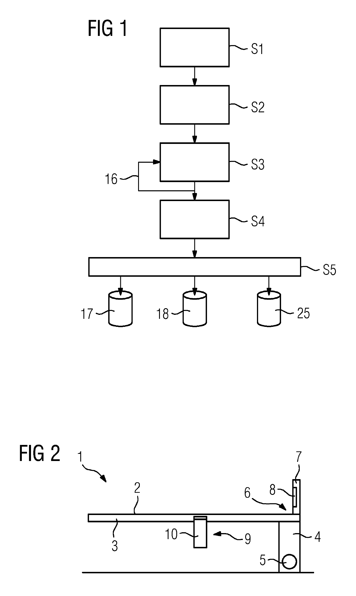
[0050]FIG. 1 depicts a flow diagram of an illustrative embodiment of the method for determination of joint load information. In an act S1, a patient to be examined is placed on a patient couch of an imaging apparatus, here an X-ray apparatus, which permits a wide variety of robot-based settings of the recording device.

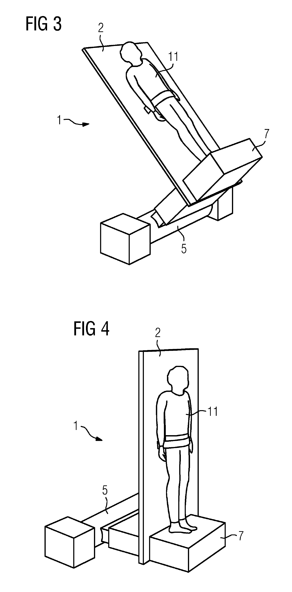
[0051]A patient couch 1 that may be used here is explained in more detail with reference to FIGS. 2 to 4.

[0052]FIG. 2 depicts a schematic sketch of the patient couch 1. The patient couch 1 has a patient table 3 with a supporting surface 2 on which the patient to be examined may be placed. The patient table 3 is carried by a stand 4, wherein it is moreover tiltable about a rotation axis 5 close to the floor, here mounted on the floor. The rotation axis 5 is arranged at the foot-side end 6 of the supporting surface 2.

[0053]At the foot-side end of the supporting surface 2, the latter is moreover terminated by a foot plate 7, which extends perpendicularly with respect to t...


PUM


Abstract

A method is provided for the automatic determination of at least one joint load information item concerning a joint of a patient, wherein at least one image data set of the loaded joint is recorded by an imaging apparatus. Several image data sets of the joint, of which at least one is three-dimensional, are recorded in each case for different loading states by the imaging apparatus. By combined evaluation of the image data sets, the at least one joint load information item is determined.

Description

[0001]The application claims the benefit of German Patent Application No. DE 10 2016 200 202.3, filed Jan. 11, 2016, which is hereby incorporated by reference in its entirety.TECHNICAL FIELD[0002]The disclosure relates to a method for the automatic determination of at least one joint load information item concerning a joint of a patient, wherein at least one image data set of the loaded joint is recorded by an imaging apparatus. The disclosure additionally relates to an imaging apparatus, such as an X-ray apparatus, a patient couch for such an imaging apparatus, and a computer program.BACKGROUND[0003]The imaging of joints, (for example of the feet, knees, hips, and spinal column), is well known in the field of medical diagnosis for the purpose of being able to establish the presence of various pathological states. It has also been proposed to record images of such joints in a weight-loaded state, e.g., in order to be able to ascertain musculoskeletal defects. It has been found that ...

Claims


Application Information

Patent Timeline
Patent Type & Authority Patents(United States)
IPC IPC(8): A61B5/05G06T7/00G06K9/00A61B6/04A61B5/00A61B5/11H04N7/18A61B6/00H04N9/47G01G19/44
CPCA61B6/505A61B5/1121A61B5/4528A61B6/0407G06K9/00362G06T7/0012A61B6/5217G06T2207/10116G01G19/44G06T2207/10012G16H50/30A61B6/0487G06V40/10
Inventor FIESELMANN, ANDREASJEREBKO, ANNA
Owner SIEMENS HEALTHCARE GMBH
Why Eureka
Try Eureka