
Method for detecting a genetic variant

a genetic variant and method technology, applied in the field of methods of detecting genetic variants, can solve the problems of high error rate of dna sequencing platforms, difficult or impossible to determine whether a genetic variant identified is real, and inaccurate determination of nucleotide positions in dna templates, etc., to achieve improved statistical confidence and high probability

Active Publication Date: 2020-05-05
CANCER RES TECH LTD
View PDF2 Cites 2 Cited by
  • Summary
  • Abstract
  • Description
  • Claims
  • Application Information

AI Technical Summary

Benefits of technology

[0032]Advantageously, the method allows detection of genetic variants at very low frequency within a DNA sample. Accordingly, the method enables earlier identification (e.g. in the context of disease pathology such as cancer-related mutations) of the presence of genetic mutations (i.e. when the mutation is present at a lower frequency) in comparison with previously known methods for mutation detection. The method therefore finds use in a variety of applications, such as screening for tumour reappearance, tumour growth, development of drug resistance / tumour evolution and identification of therapeutically actionable mutations. The method also allows for the identification of the presence of genetic mutations at low frequencies with improved statistical confidence.
[0073]Many of the above samples can be obtained non-invasively, and can therefore be taken regularly without great risk or discomfort to the subject.

Problems solved by technology

DNA sequencing platforms have error rates.
For example, occasionally the polymerase used in the amplification reaction will incorporate the wrong nucleotide base in the complementary strand being synthesised, leading to an incorrect determination of the nucleotide at that position in the DNA template.
This is problematic especially for the detection of genetic variants that will only be present in a DNA sample at low frequency, for example a frequency approaching or below the error rate of the sequencing method used.
Under such circumstances, it is difficult or impossible to determine whether a genetic variant identified is real (i.e. actually present in the DNA template molecule) or an error.
Therefore, detecting mutations which are present in a DNA sample at frequency of ˜1% or lower is problematic.
Existing methods of DNA sequencing and genetic variant identification have limitations with regards to the detection of rare, novel variants in multiple regions, especially in samples having small amounts of DNA.
Methods are typically incapable of identifying mutations occurring at a frequency lower than or similar to the error rate of method used (i.e. background noise).
Moreover, dPCR has a limited scope for analysing multiple regions of interest in parallel, especially where DNA sample is limited.
These methods are not suitable for simultaneous analysis of multiple genes (i.e. multiple regions of interest) and when DNA is limited.
However, major challenges persist in this field, such as (a) screening sufficient bases of the genome to detect relevant cancer mutations (b) screening of small quantities of fragmented DNA for such mutations, and (c) detection of low frequency mutant tumour DNA molecules amongst many ‘wild-type’ molecules.

Method used


Image

  • Method for detecting a genetic variant
  • Method for detecting a genetic variant
  • Method for detecting a genetic variant

Examples


example 1

Determination of Background Noise

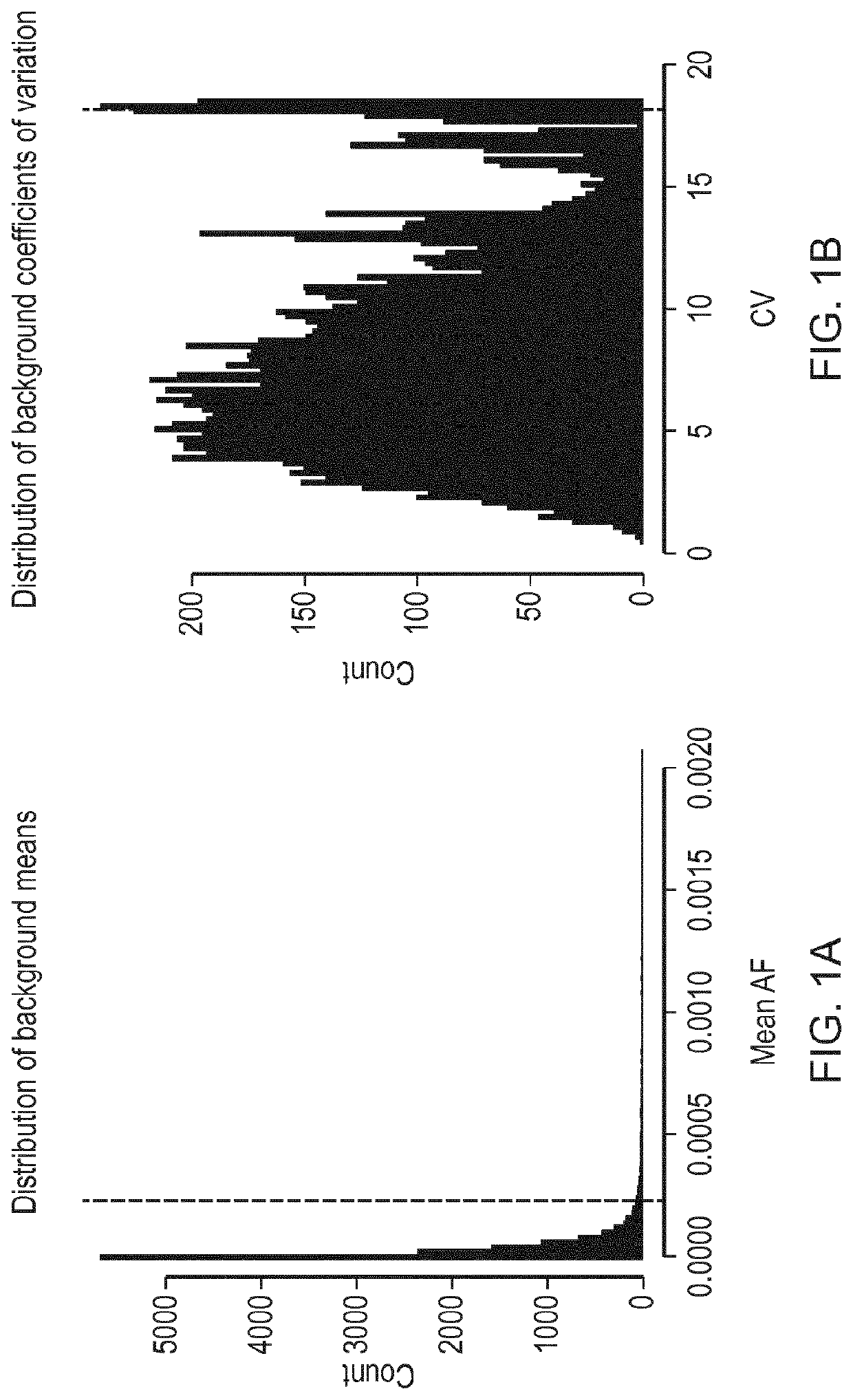
[0188]41 amplicons were designed, covering all of TP53 and hotspot regions in EGFR, KRAS, BRAF and PIK3CA, and were read in forward and reverse directions, giving 82 read families in total (Table 1). The amplicons cover 5,038 bases including overlapping forward and reverse reads (excluding primer sequences).

[0189]

TABLE 1LeftRightChromosomecoordinatecoordinateAmplicon namechr7140453098140453187BRAF_D0016_001_Fchr7140453128140453217BRAF_D0016_001_Rchr75524158955241678EGFR_D0016_001_Fchr75524162055241709EGFR_D0016_001_Rchr75524165855241745EGFR_D0016_002_Fchr75524165955241746EGFR_D0016_002_Rchr75524170555241792EGFR_D0016_003_Fchr75524170655241793EGFR_D0016_003_Rchr75524238555242474EGFR_D0016_004_Fchr75524239755242486EGFR_D0016_004_Rchr75524242755242516EGFR_D0016_005_Fchr75524244855242537EGFR_D0016_005_Rchr75524893155249020EGFR_D0016_006_Fchr75524893855249027EGFR_D0016_006_Rchr75524898955249078EGFR_D0016_007_Fchr75524901155249100EGFR_D0016_007_Rchr7552490...

example 2

Determining the AF Required for a Positive Call for a Base Change in a Given Reaction

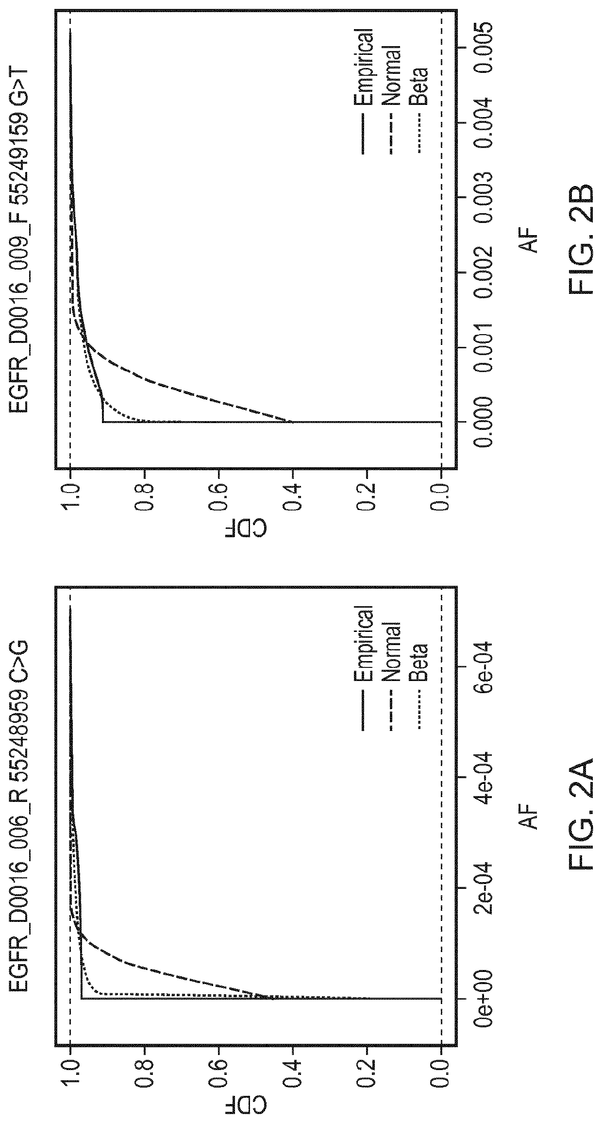
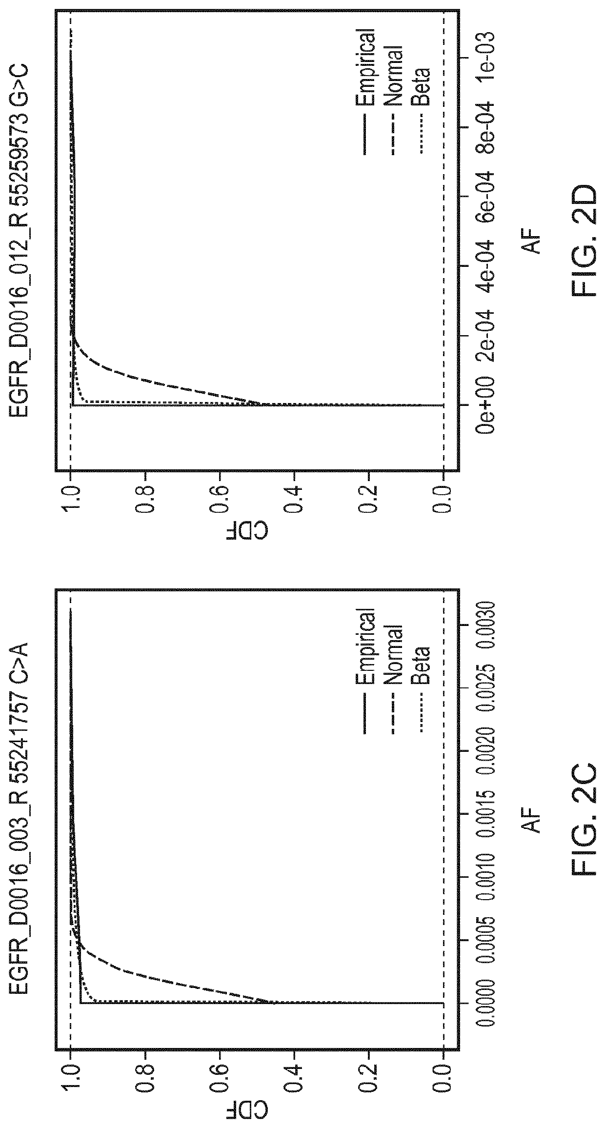
[0195]A reaction was defined as positive for a given sequence change if the observed AF was found to be greater than the minimal detectable frequency (MDF) for that allele. The MDF for an allele was calculated as the AF at which the cumulative distribution function (CDF) for that particular base change crosses a predefined threshold (CDF_thresh).

[0196]FIG. 2 shows cumulative distributions for several base changes of the panel of Example 1. Cumulative distribution functions for the Normal and Beta distributions are shown, fitted using the values for the mean AF and CV that were calculated for those base changes, compared against the empirical distributions of the data. The Kolmogorov-Smirnov value for goodness of fit for the Normal (KS

[0197]Normal) and Beta (KS Beta) distributions to the empirical distributions of the data are as follows:

[0198]

Base changeKS NormalKS BetaEGFR_D0016_006_R 55248959 C > ...

example 3

Determining the Number of Replicate Amplification Reactions to be Positive for a Given Base Change to Determine Presence of the Base Change in a Sample

[0205]To minimise false positive determinations for the presence of a base change in a sample, multiple positive replicate amplification reactions (N) were required out of the total number of replicate amplification reactions for that sample (T).

[0206]The theoretical probability of a false positive appearing in N out of T reactions (P_fpNT) is

P_fpNT=(P_fp1T){circumflex over ( )}N)*C_NT=((1−CDF_thresh){circumflex over ( )}N)*C_NT

where “C_NT” is the number of possibilities of choosing N wells out of T without attention to order, and is

C_NT=T! / ((T−N)!*N!)

where “!” indicates the factorial function.

[0207]For example, for choosing 3 reactions out of 48 there are 17,296 possibilities. For choosing 2 reactions out of 48 there are 1,128 possibilities. The expected rate of false positives was calculated for each sample and is shown in Tables 2...


PUM

PropertyMeasurementUnit
threshold frequencyaaaaaaaaaa
frequencyaaaaaaaaaa
threshold frequenciesaaaaaaaaaa

Abstract

The present invention provides a method for detecting a genetic variant in a region of interest in a DNA sample comprising (i) determining, for a given sequencing platform, sequencing process and sequencing depth, the distribution of the number of reads supporting a genetic variant or plurality of genetic variants expected to be observed in the sequencing results of amplification reactions due to amplification and sequencing error (read count distribution); (ii) based on the read count distribution determined in step (i), establishing a threshold frequency at or above which the genetic variant must be observed in sequencing results of amplification reactions to assign a positive determination for the presence of the genetic variant in a given amplification reaction; (iii) partitioning the DNA sample into a plurality of replicate amplification reactions, so that the mean number of amplifiable template molecules of the region of interest in a replicate amplification reaction is fewer than the reciprocal of the threshold frequency determined in step (ii); (iv) performing the amplification reactions of step (iii) and sequencing the products of amplification reactions, (v) based on step (ii) and the results of step (iv), determining the presence / absence of the genetic variant in each replicate amplification reaction; and (vi) integrating the results of (v) to determine the presence / absence of the genetic variant in the region of interest in the DNA sample.

Description

FIELD OF THE INVENTION[0001]The present invention relates to methods of detecting genetic variants in DNA samples.BACKGROUND TO THE INVENTION[0002]A genetic variant is one or more nucleotides which differ from a reference DNA sequence for a given region. For example, a genetic variant may comprise a deletion, substitution or insertion of one or more nucleotides.[0003]A DNA sample may be analysed for known genetic variants or to discover previously unknown genetic variants in a region of interest by determining the DNA sequence in the region of interest and comparing the determined sequence to the reference sequence.[0004]DNA sequencing can be performed using a variety of techniques, such as the classic chain termination method, or one of several high-throughput, next generation sequencing (NGS) methodologies, reviewed by Metzker, M. L., Nat Rev Genet 2010 January; 11(1): 31-46.[0005]Illumina sequencing, 454 pyrosequencing, Heliscope single molecule sequencing, single molecule real t...

Claims


Application Information

Patent Timeline
Patent Type & Authority Patents(United States)
IPC IPC(8): C12Q1/6858G16B20/00C12Q1/6827C12Q1/6886C12Q1/6883G16B20/20
CPCC12Q1/6886C12Q2545/114G16B20/00C12Q1/6858C12Q2535/122C12Q1/6883C12Q1/6827C12Q2563/159C12Q2537/165C12Q2600/156C12Q2600/106C12Q2527/146C12Q2600/118G16B20/20G16B30/00C12Q1/6869C12Q2535/00
Inventor ROSENFELD, NITZANFORSHEW, TIMMARASS, FRANCESCOMURTAZA, MUHAMMED
Owner CANCER RES TECH LTD
Learn More