AAV-IDUA vector for treatment of MPS I-associated blindness
a technology of aavidua and aav, applied in the field of viral vectors, can solve the problems mps i-associated blindness, and common deficiency of hsct and aav cns-targeted or systemic gene therapy, and achieve the effect of correcting mps i-associated blindness in privileged
- Summary
- Abstract
- Description
- Claims
- Application Information
AI Technical Summary
Problems solved by technology
Method used
Image
Examples
example 1
Materials and Methods
[0145]Production of AAV vectors. For cell culture experiments, a previously described triple transfection method was used to generate the vectors used herein (Grieger et al., Nat. Protoc. 1(3):1412 (2006)). This method used the pXR2 or pXR8G9 plasmids, which all contain rep2 of AAV and individually the capsid genes of the indicated serotype. The plasmid containing the intended AAV genome was first constructed by substituting the egfp gene in pTR-CMV-eGFP with the codon optimized IDUA cDNA (provided by GenScript) at the AgeI and SalI sites. Following AAV production and cesium chloride gradient separation (Grieger et al., Nat. Protoc. 1(3):1412 (2006)), peak fractions were dialyzed against PBS, and titered by quantitative PCR (CMV_F CAA GTA CGC CCC CTA TTG AC (SEQ ID NO:2), CMV_R AAG TCC CGT TGA TTT TGG TG (SEQ ID NO:3)) which was confirmed by Southern dot blot (Grieger et al., Nat. Protoc. 1(3):1412 (2006)). For the experiments in human explants, GMP grade vector...
example 2
AAV IDUA Expression Cassette in MPS I Fibroblasts
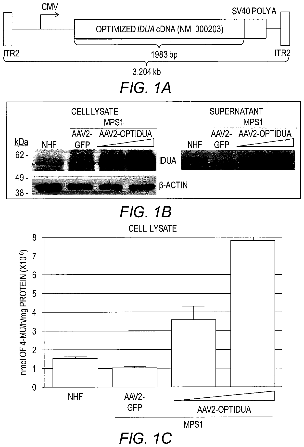
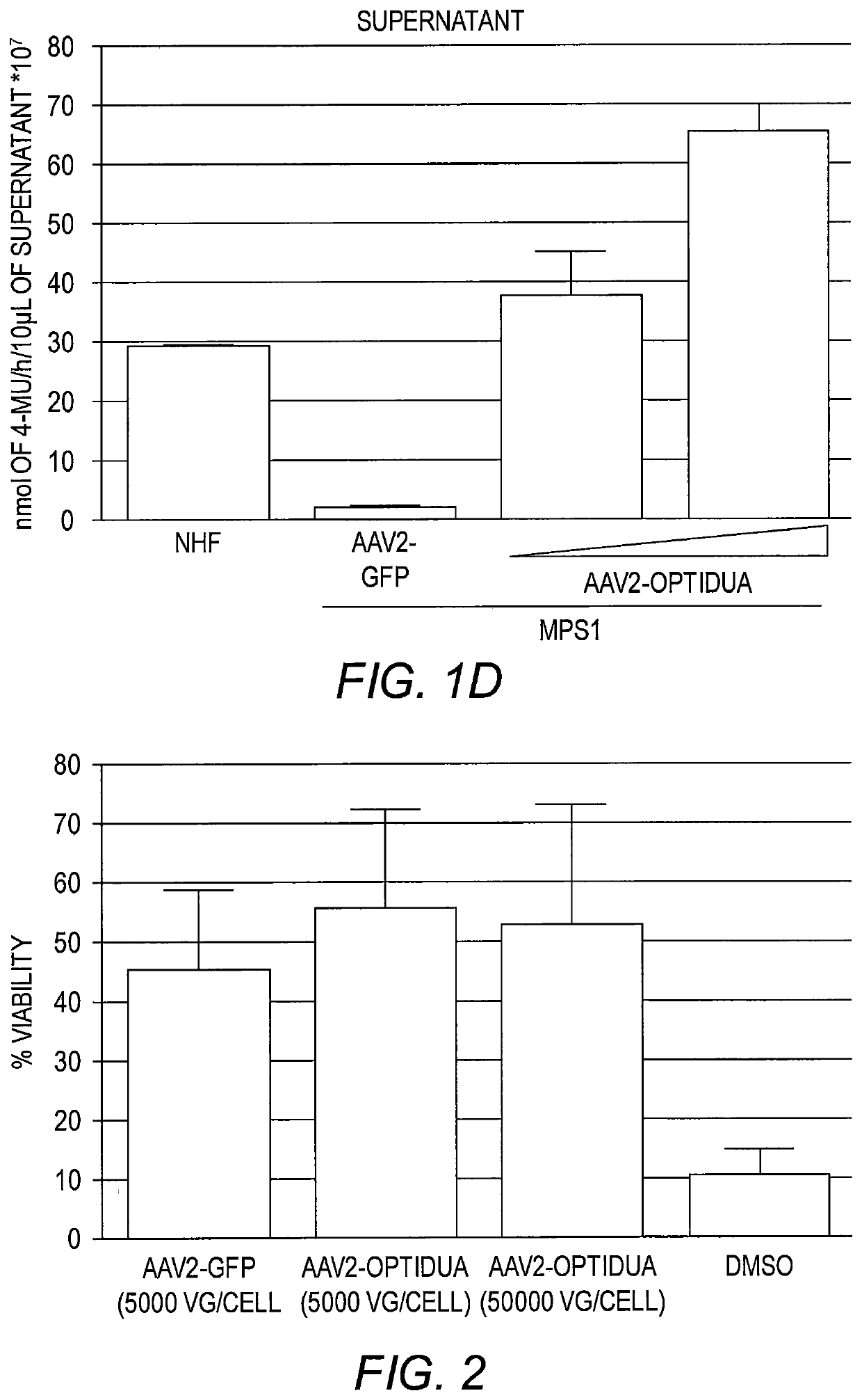
[0155]To develop an AAV IDUA expression cassette, the human idua cDNA (NM_000203) was codon optimized for human expression (opt-IDUA) and situated between the CMV promoter and the SV40 poly-adenylation sequence in an AAV inverted terminal repeat serotype 2 plasmid context (FIG. 1A). AAV2-opt-IDUA vectors were prepared as described (Grieger et al., Adv. Biochem. Eng. Biotechnol. 99:119 (2005)) and used for characterization in MPS I patient derived fibroblasts. Immortalized normal human fibroblasts (NHF) served as the control cell line (Simpson et al., J. Carcinog. 4:18 (2005)). In dose escalation experiments, AAV2-opt-IDUA transduction of MPS I fibroblasts resulted in increasing levels of IDUA restoration, in both cellular lysates and in the culture supernatant (FIG. 1B). In fact, as resting levels of IDUA in NHF are relatively low, a dose of 5,000 viral genomes / cell resulted in already supraphysiological levels in both cell lysates an...
example 3
AAV IDUA Expression Cassette in Corneas
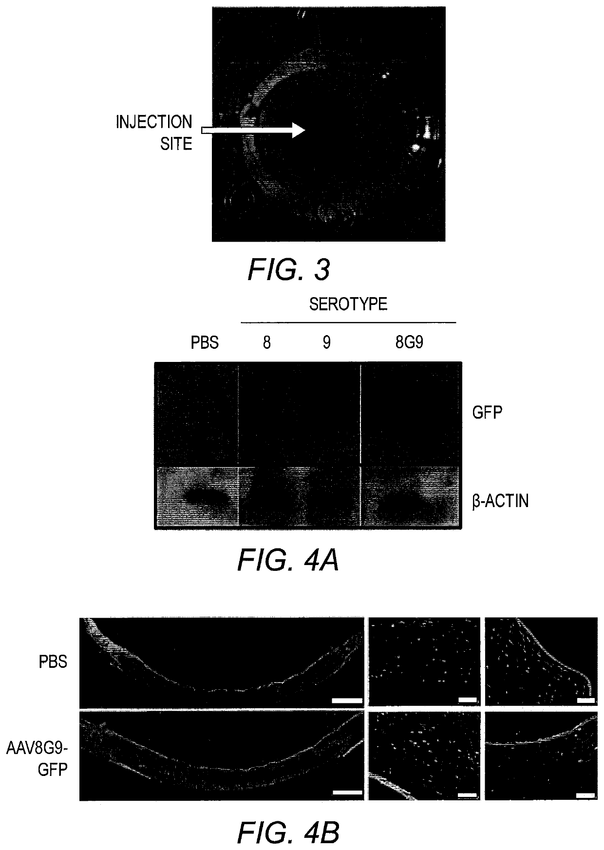
[0156]An analysis of MPS I patient corneas attributed stromal abnormalities as the cause of the corneal opacity that results in vision loss (Huang et al., Exp. Eye Res. 62(4):377 (1996)). Therefore, direct administration of AAV-opt-IDUA should restore IDUA activity in the corneal stroma compartment of MPS I patients, hopefully preventing or reversing the MPS I phenotype. Previous reports demonstrated the utility of both AAV8 (Hippert et al., PLoS One, 7(4):e35318 (2012)) and AAV9 (Sharma et al., Exp. Eye Res. 91(3):440 (2010)) for human keratocyte transduction, one of the most prevalent cell types present in the cornea. As such, these capsids, carrying a self-complementary (sc) AAV-CMV-GFP genome, were evaluated in normal human corneal explants, which remain viable for weeks post-mortem. Preliminary experiments demonstrated that a volume of 50 μl injected into the stroma was tolerated with minimal tissue distension and a distribution across ≈50...
PUM
| Property | Measurement | Unit |
|---|---|---|
| pH | aaaaa | aaaaa |
| thickness | aaaaa | aaaaa |
| volume | aaaaa | aaaaa |
Abstract
Description
Claims
Application Information
 - R&D Engineer
- R&D Manager
- IP Professional
- Industry Leading Data Capabilities
- Powerful AI technology
- Patent DNA Extraction
Browse by: Latest US Patents, China's latest patents, Technical Efficacy Thesaurus, Application Domain, Technology Topic, Popular Technical Reports.
© 2024 PatSnap. All rights reserved.Legal|Privacy policy|Modern Slavery Act Transparency Statement|Sitemap|About US| Contact US: help@patsnap.com


