Methods and systems for monitoring cell exocytosis or endocytosis

a cell exocytosis and cell technology, applied in the field of methods and systems for monitoring cell exocytosis or endocytosis, can solve the problems of slow and specificity, inability to easily apply single cells, and high cost of reagent use, and achieve the effect of cheap and easy to implemen

Inactive Publication Date: 2005-01-13
MEDICAL RESEARCH COUNCIL
View PDF4 Cites 4 Cited by
  • Summary
  • Abstract
  • Description
  • Claims
  • Application Information

AI Technical Summary

Benefits of technology

[0013] Application of IRM to the study of stimulated or inhibited secretion, or to the study of small organelles, provides a new approach that can be relatively cheap and easy to implement but can also be used in conjunction with electrophysiological techniques, such as capacitance measurements, and fluorescence imaging techniques, such as confocal microscopy and TIRFM. IRM can overcome two major drawbacks of the capacitance technique. First, exocytosis and endocytosis can be investigated while voltage-sensitive conductances are active because the cell does not need to be voltage-clamped. We have taken advantage of this to measure exocytosis in bipolar cells stimulated by current injection and demonstrate that exocytosis during normal electrical activity occurs at rates at least 10-fold slower than the maximum. Second, measurements can be made from neurons with small synaptic terminals and complex morphologies. We have taken advantage of this to investigate exocytosis and endocytosis in the synaptic terminal of bipolar cells from the mouse retina. The visualization of fusion by dense-core granules may also make IRM a useful method for studying Ca2+-triggered secretion in cell-free systems (Avery et al., 2000; Holroyd et al., 2002).
[0014] Further advantages of the IRM approach are that it is applicable to a wide-range of cell types and is independent of the substance(s) being secreted. It is also non-invasive, does not induce photodamage, and does not require a skilled operator.
[0037] The present invention also has particular utility as a screening assay. For example, the effect a bioactive agent may have on the exocytic or endocytic response of a cell can be tested by introducing that bioactive agent to the cell. The introduced bioactive agent may then directly stimulate or inhibit secretion, or the bioactive agent may have an effect on secretion stimulated or inhibited in other ways (e.g. electrically, or by changing the chemical environment of the cell). Whichever way the change in the amount of secretion is provoked, the secretion can be detected by analysis of the IRM light signals. Such analysis will generally be much quicker to complete than e.g. biochemical assays (which attempt to detect the chemical that is released). The speed at which the signals can be obtained and analysed makes high throughput screening assays based on the method highly attractive. Also, in contrast, to biochemical assays, the method is not specific to particular secreted substances, but has more general applicability.
[0059] An advantage of the apparatus is that the signal analyser automatically detects exocytosis or endocytosis, thereby making the apparatus particularly suitable for use in high throughput screening of bioactive agents or the screening of cell populations. Preferably the signal analyser is adapted to determine the amount of exocytosis or endocytosis at the membrane.

Problems solved by technology

Analytical methods rely on detecting the substance released, rather than the process of secretion itself, and are therefore slow and specific to the chemical being detected.
They are not easily applied to single cells and are costly in the use of reagents.
A second drawback of the capacitance technique is that it cannot monitor exocytosis during normal electrical activity because voltage-sensitive conductances, such as sodium and calcium channels, can generate spurious signals (Horrigan and Bookman, 1994; Gillis, 1995).
Further, the capacitance technique is invasive; the properties of exocytosis and endocytosis can be severly altered by the dialysis of the cell interior through the whole-cell pipette.
A major drawback of amperometry is that only some secreted substances can be detected.
These imaging methods generally have lower time-resolution than the capacitance technique.
Toxic photodamage induced by the intense lights required to excite fluorescence is an important drawback and limitation of these methods.

Method used


Image

  • Methods and systems for monitoring cell exocytosis or endocytosis
  • Methods and systems for monitoring cell exocytosis or endocytosis
  • Methods and systems for monitoring cell exocytosis or endocytosis

Examples


Embodiment Construction

[0099] Experimental Procedures

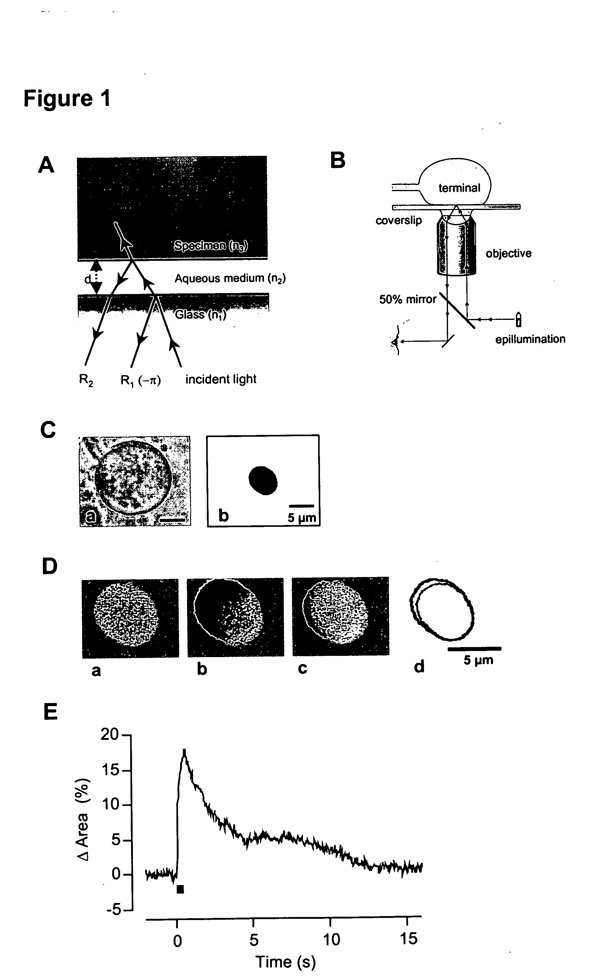
[0100] Interference Reflection Microscopy

[0101] The theory of image formation has been described in detail by Izzard and Lochner (1976) and Verschueren (1985). The method depends on the fact that light is reflected at the interface between media of different refractive indices. When a cell on a coverslip is observed under epi-illumination using an oil-immersion objective, the first two interfaces where the light is reflected are the glass-solution and the solution-cell boundaries (FIG. 1A). If the thickness of the aqueous solution is of the order of the wavelength of light used (λ), the two reflected beams, R1 and R2, interfere. The optical path difference (Δ) between R1 and R2 depends on the distance between the interfaces (d). If d=0, Δ=λ / 2, and there is maximum destructive interference. The two beams are half a wavelength out of phase because R1 is reflected at the interface between a medium of high refractive index (glass, n1) and lower refractive...


PUM

PropertyMeasurementUnit
diameteraaaaaaaaaa
diametersaaaaaaaaaa
diametersaaaaaaaaaa

Abstract

A method of assaying exocytosis or endocytosis, comprises: providing a transparent substrate with at least one cell membrane mounted thereon apposing the substrate, provoking a change in the amount of exocytosis or endocytosis at the membrane, obtaining light signals from the region of the substrate which the membrane apposes using interference reflection microscopy, and analysing the light signals to detect the exocytosis or endocytosis at the membrane.

Description

CROSS-REFERENCE TO RELATED APPLICATIONS [0001] This application claims priority under 35 USC §119(e) to U.S. Provisional Patent Application 60 / 485,538 filed 8 Jul. 2003, the entirety of which is incorporated by reference herein.FIELD OF THE INVENTION [0002] The present invention relates to methods and systems for monitoring cell exocytosis or endocytosis using interference reflection microscopy (IRM). BACKGROUND OF THE INVENTION [0003] The traffic of secretory vesicles to the plasma membrane of eukayrotic cells is essential for normal cellular function. It also forms the basis of intercellular communication in multicellular organisms through the release of a wide array of extracellularly acting molecules. The process of secretion is generally classified into two types: regulated and constitutive (Burgoyne and Morgan, 2002). Regulated secretion takes place when a vesicle or granule rapidly fuses with the surface membrane (exocytosis) in response to an external stimulus, releasing its...

Claims


Application Information

Patent Timeline
Patent Type & Authority Applications(United States)
IPC IPC(8): C12M1/34C12Q1/00G01N33/50G01N33/543G02B21/33
CPCG01N33/502G02B21/33G01N33/54373
Inventor LAGNADO, LEONLLOBET, ARTUR
Owner MEDICAL RESEARCH COUNCIL
Why Eureka
Try Eureka