Method of segmenting a radiographic image into diagnostically relevant and diagnostically irrelevant regions

a radiographic image and segmentation technology, applied in image enhancement, medical/anatomical pattern recognition, instruments, etc., can solve the problems of not working well, method cannot deal with single-exposure radiographs, and cannot be used to completely segment diagnostically relevant regions

Active Publication Date: 2005-01-27
CARESTREAM HEALTH INC
View PDF8 Cites 31 Cited by
  • Summary
  • Abstract
  • Description
  • Claims
  • Application Information

AI Technical Summary

Benefits of technology

The invention has the following advantages. 1. It is independent of the exam type of the radiograph. 2. It can automatically segment a radiograph into foreground, background and

Problems solved by technology

The thresholds used in this method also vary from one exam-type (bodypart and projection) to the other, which may not work well when exam-type information is unavailable.
However, this method can only deal with single-exposed radiographs, and can not be used to completely segment the diagnostically

Method used


Image

  • Method of segmenting a radiographic image into diagnostically relevant and diagnostically irrelevant regions
  • Method of segmenting a radiographic image into diagnostically relevant and diagnostically irrelevant regions
  • Method of segmenting a radiographic image into diagnostically relevant and diagnostically irrelevant regions

Examples


Embodiment Construction

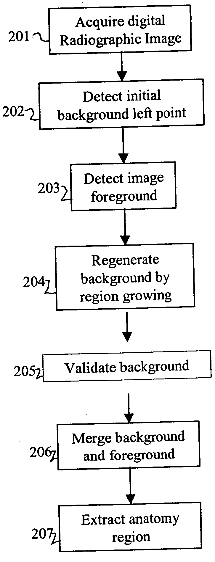
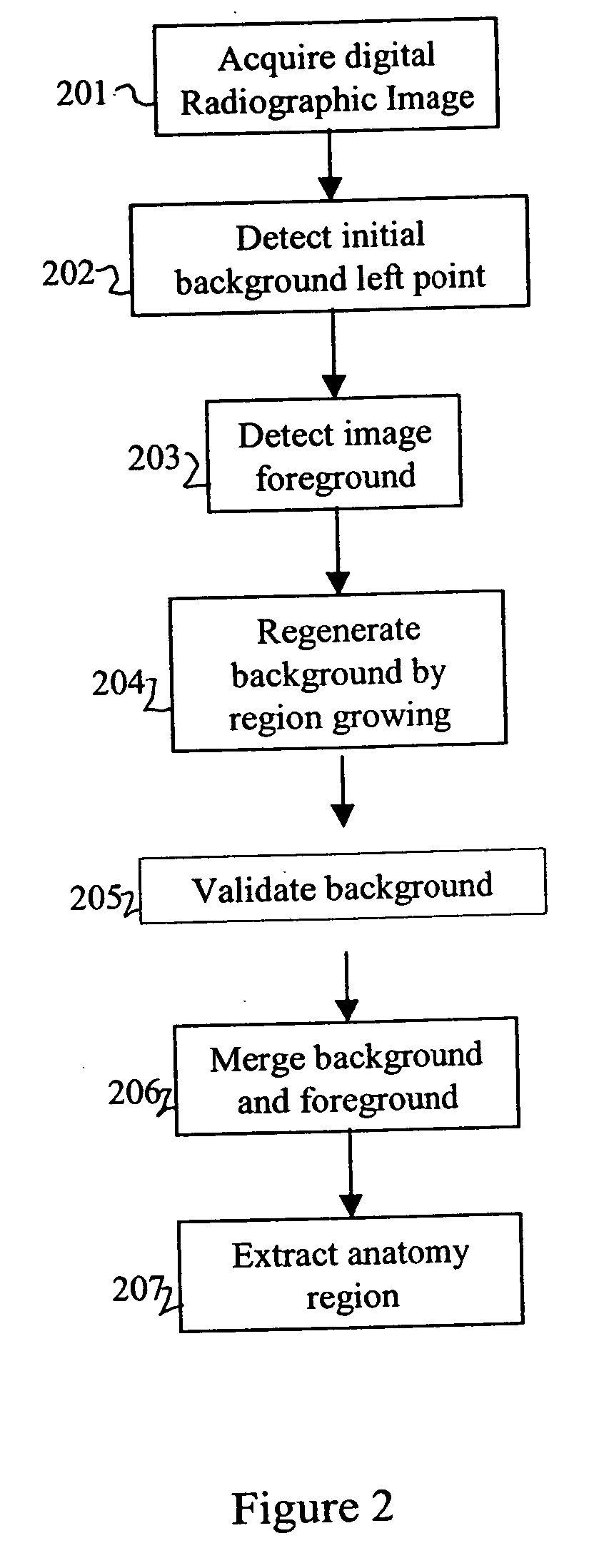
Referring now FIG. 2, an embodiment of the present invention will be described.

A digital radiographic (medical) image is acquired (box 201) such as from a diagnostic imaging device (MRI, CT, PET, US), from a direct digital or computed radiography device, from an x-ray film digitizer, from a medical image archive. The digital medical image includes an array or matrix of rows (lines) and columns of pixels having a gray scale range of a predetermined range of bits or code values (e.g., 8 or 12 bits). The digital medical image is processed in an image processor, such as a digital computer and output to a display or hard copy. The method of the invention is carried out in the image processor.

This invention first tries to improve the background detection algorithm disclosed in U.S. Pat. No. 5,606,587, issued Feb. 25, 1997, inventors Barksi et al., by making it exam-type independent and at the same time to provide a reasonable initial background left point (box 202) for the acquired d...


PUM


Abstract

A method of segmenting a radiographic image into diagnostically relevant and diagnostically irrelevant regions comprising: acquiring a digital radiographic image including a matrix of rows and columns of pixels; detecting the initial background left point of a histogram of the image; detecting the foreground of the image; regenerating the background of the image by region growing; validating the background of the image; merging the background and foreground regions of the image as diagnostically irrelevant regions; and extracting the anatomy region of the image as the diagnostically relevant region.

Description

FIELD OF THE INVENTION This invention relates in general to the processing of digital radiography, and in particular to the segmentation of a digital radiographic image into diagnostically relevant and diagnostically irrelevant regions. BACKGROUND OF THE INVENTION The recent advance in digital radiographic imaging systems, such as the flat-panel based direct digital radiographic (DR) systems (“A high resolution, high frame-rate, flat-panel TFT array for digital x-ray imaging,” Proceedings of SPIE Medical Imaging, Antonuk et al., vol. 2163, pp 118-128, 1994) and the storage phosphor based computed radiographic (CR) systems (“Introduction to medical radiographic imaging,” Eastman Kodak Company, 1993) etc, allow the separation and optimization of image acquisition, processing, and display processes individually. The image pixel data are manipulated during the image-processing step so as to optimize the image information acquired on the radiograph and to help the radiologists to bette...

Claims


Application Information

Patent Timeline
IPC IPC(8): G06T1/00G06T5/00G06T5/40A61B6/00G06T7/00G06T7/60G06V10/28
CPCG06K9/342G06K9/38G06K2209/05G06T2207/30004G06T2207/10116G06T2207/20061G06T7/0081G06T7/11G06V10/28G06V10/267G06V2201/03
Inventor WANG, XIAOHUILUO, HUI
Owner CARESTREAM HEALTH INC
Why Eureka
Try Eureka