Eyeground image blood vessel segmentation method based on self-adaption difference of Gaussians

A fundus image and Gaussian difference technology, which is applied in the field of biomedical image processing, can solve the problems of insensitivity to brightness and contrast, and achieve the effect of suppressing the influence of blood vessel segmentation.

Active Publication Date: 2015-09-09
SHANGHAI NEW EYES MEDICAL CO LTD
View PDF3 Cites 15 Cited by
  • Summary
  • Abstract
  • Description
  • Claims
  • Application Information

AI Technical Summary

Problems solved by technology

[0004] The purpose of the present invention is to overcome the above-mentioned deficiencies in the prior art, and proposes a method fo

Method used


Image

  • Eyeground image blood vessel segmentation method based on self-adaption difference of Gaussians
  • Eyeground image blood vessel segmentation method based on self-adaption difference of Gaussians
  • Eyeground image blood vessel segmentation method based on self-adaption difference of Gaussians

Examples


Embodiment Construction

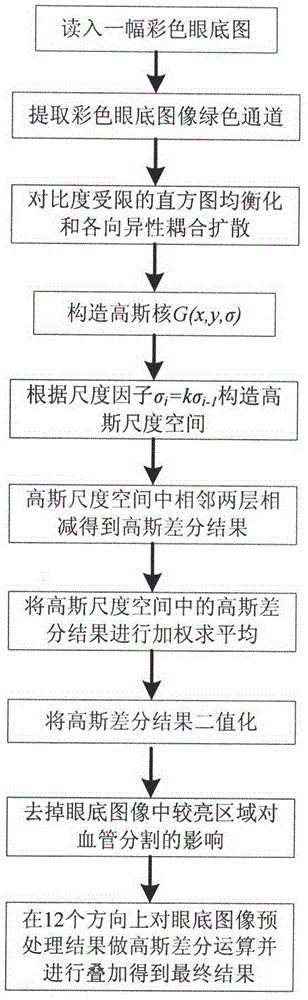
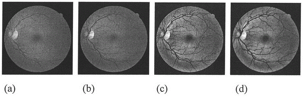
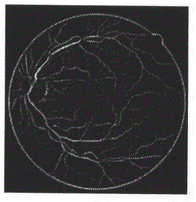
[0036] The flow chart of the present invention is as figure 1 As shown, the green channel of the fundus image is firstly extracted, and the contrast of the image is improved by using a contrast-limited adaptive histogram equalization; the anisotropic coupling diffusion equation is used for filtering to improve the definition of blood vessels; and then the adaptive Gaussian based The difference algorithm performs blood vessel segmentation on the fundus image; and the blood vessels of the Gaussian difference result are enhanced Figure II value, remove the influence of bright areas on the blood vessel segmentation results; finally superimpose the segmentation results of 12 directions to get the final result, to ensure that the blood vessels in each direction are detected. The specific implementation process of the technical solution of the present invention will be described below in conjunction with the accompanying drawings.

[0037] 1. Extract the green channel G(x, y) of th...


PUM


Abstract

The invention discloses an eyeground image blood vessel segmentation method based on self-adaption difference of Gaussians. The segmentation method comprises steps of 1) extracting colorful eyeground image green channels, and performing pretreatment of self-adaption histogram equalization and anisotropy coupling diffusion with limited contrast ratio; 2) constructing Gaussian scale space; 3) subtracting adjacent two layers in the Gaussians scale space so as to get difference of Gaussian images; 4) averaging difference of Gaussian image weighing so as to get blood vessel increasing images; 5) performing binaryzation for the blood vessel increasing images; 6) rotating Gaussian kernel in 12 directions, wherein 15 DEG is regarded as step size in 0-180 DEG, repeating steps of 2-5, and overlapping results in the 12 directions; 7) selecting 20% of the grey value of the second peak as a light area of a threshold value extracting images according to bimodality of a pretreatment image histogram; and 8) reducing light areas from the blood vessel binary images so as to reduce effects of the light areas on segmentation of blood vessels. The method is widely applicable for blood vessel segmenetaion of all kinds of colorful eyeground images.

Description

technical field [0001] The invention belongs to the technical field of biomedical image processing, and relates to a method for segmenting blood vessels in fundus images based on self-adaptive Gaussian difference, which can be used for segmenting blood vessels in fundus images with different degrees of normal and pathological changes. Background technique [0002] Changes in fundus vascular structure are symptoms of many diseases such as diabetes, hypertension, cardiovascular disease, and stroke. Among them, changes in vessel diameter, bifurcation angle, and vessel distortion are symptoms of hypertension, and the formation of new blood vessels is a sign of diabetes. , In developing countries, complications of diabetes are the cause of blindness in the eyes, and local thinning of arteriovenous vessels is an important precursor of stroke. Early detection of these vascular changes in the fundus is very important for doctors to perform early intervention on patients and prevent ...

Claims


Application Information

Patent Timeline
IPC IPC(8): G06T7/00G06T7/40
CPCG06T7/12G06T7/44G06T2207/20004G06T2207/30041G06T2207/30101
Inventor 肖志涛张芳李敏耿磊吴骏张欣鹏杜伟强
Owner SHANGHAI NEW EYES MEDICAL CO LTD
Why Eureka
Try Eureka