Method and system for composite gating using multiple inputs

a composite gating and input technology, applied in the field of overall motion measurement, can solve the problems of incomplete reconstruction images, various motion related artifacts or discontinuities, and images that still possess motion artifacts

Active Publication Date: 2005-05-26
GE MEDICAL SYST GLOBAL TECH CO LLC
View PDF31 Cites 48 Cited by
  • Summary
  • Abstract
  • Description
  • Claims
  • Application Information

AI Technical Summary

Benefits of technology

[0009] In accordance with one aspect of the present invention, a technique is provided for imaging an organ. In view of this aspect, a set of motion data for an organ of interest may be acquired from at least one or more non-electrical sensors. The set of motion data may be processed to extract two or more prospective gating points and two or more retrospective gating points for the organ of interest. A set of image data representative of the organ of interest may be acquired using the two or more prospective gating points. A portion of the set of image data may be processed based upon the two or more retrospective gating points.
[0010] In accordance with another aspect of the present invention, a technique is provided for imaging an organ. In view of this aspect, a set of motion data for an organ of interest may be acquired from at least one or more non-electrical sensors. The set of motion data may be processed to extract two or more prospective gating points and two or more retrospective gating points for an organ of interest. A set of image data representative of the organ of interest may be acquired using the two or more prospective gating points. The set of image data may be reconstructed to generate a set of reconstructed data. A portion of the set of reconstructed data may be processed based upon the two or more retrospective gating points.
[0011] In accordance with one aspect of the present invention, a technique is provided for imaging an organ. In view of this aspect, a set of motion data for a respiratory organ of interest may be acquired from at least one or more electrical sensors. The set of motion data may be processed to extract two or more prospective gating points and two or more retrospective gating points for the organ of interest. A set of image data representative of the organ of interest may be acquired using the two or more prospective gating points. A portion of the set of image data may be processed based upon the two or more retrospective gating points.
[0012] In accordance with another aspect of the present invention, a tech

Problems solved by technology

The reconstructed images, however, may be flawed or contain artifacts due to the motion of internal organs, such as the heart, lungs, diaphragm, stomach, and so forth.
In particular, if the imaged region has undergone motion during the imaging process, various motion related artifacts or discontinuities may be present in the reconstructed image.
However, due to the spatial relationship of the various internal organs, many of which unde

Method used


Image

  • Method and system for composite gating using multiple inputs
  • Method and system for composite gating using multiple inputs
  • Method and system for composite gating using multiple inputs

Examples


Embodiment Construction

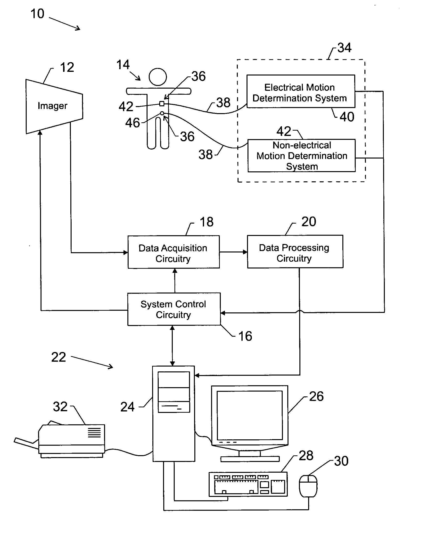
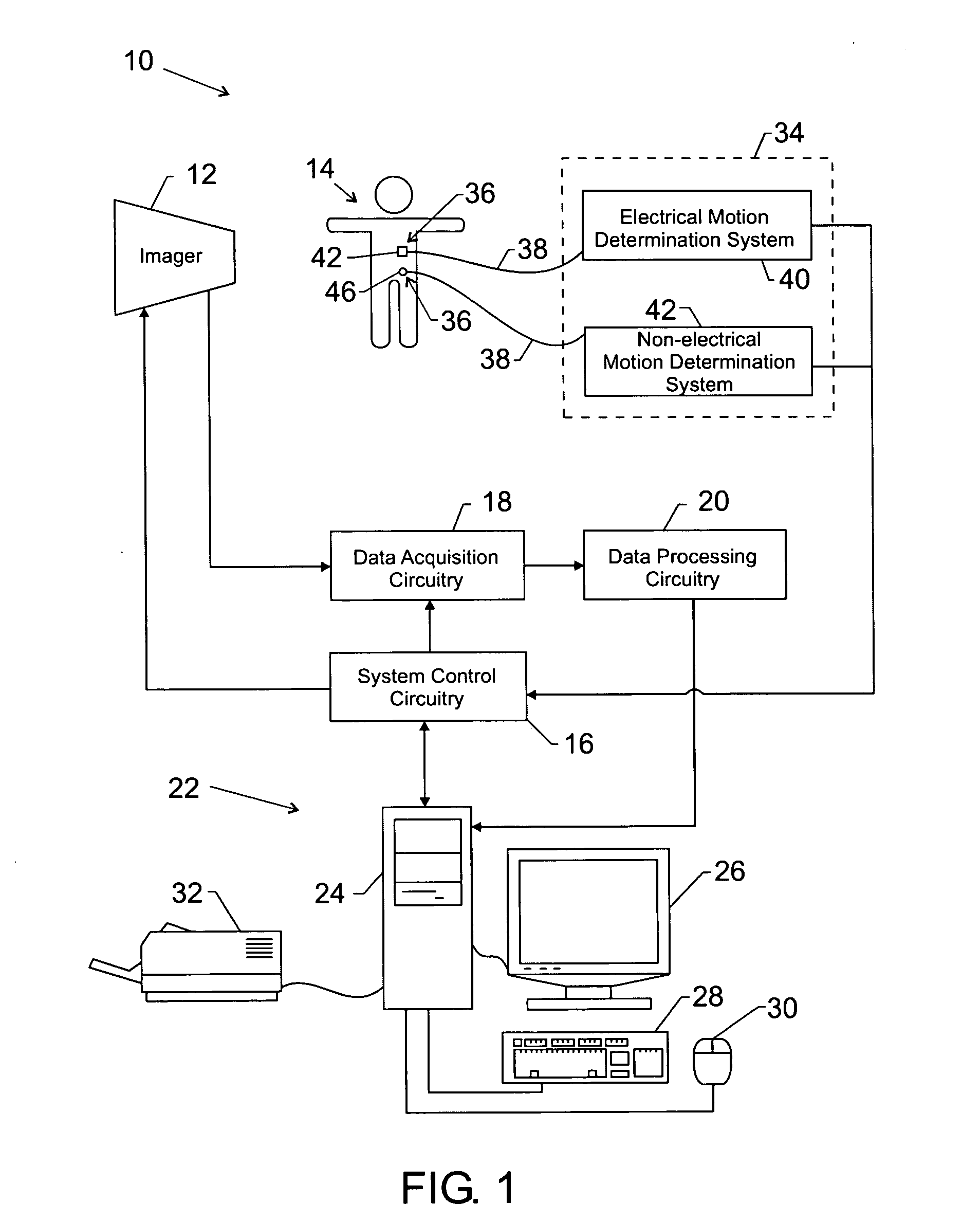
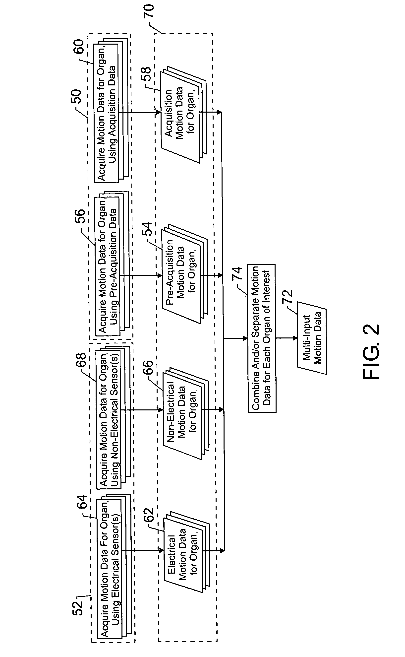
[0025] In the field of medical imaging, it is often desirable to characterize the motion of an imaged organ so that the motion may be accounted for to improve image quality. Measures of the muscular activity of the organ of interest, however, may characterize only a portion of the overall motion of the organ relative to the viewer or imaging scanner. In particular, the overall motion may consist of not only the muscular contractions of the organ of interest itself, i.e., the inherent motion, but also motion attributable to the movement of proximate organs. For example, the overall motion of the heart may consist of not only the muscular contractions of the heart but also the movement of the lungs, the movement of the diaphragm, or other proximate muscular contractions, voluntary and / or involuntary, of the patient's body. Therefore, in the example of cardiac imaging, it may be desirable to characterize the overall motion of the heart relative to the imager, not simply the motion attr...


PUM


Abstract

One or more techniques are provided for determining the overall motion of an organ of interest relative to a viewer or imager. Motion data is acquired for the organ of interest and/or for one or more proximate organs using sensor-based and/or image data-based techniques. The sensor-based techniques may include electrical and non-electrical techniques. The image data-based techniques may include both pre-acquisition and acquisition image data. The motion data for the organ of interest and proximate organs may be used to determine one or more quiescent periods corresponding to intervals of minimal motion for the organ of interest and the proximate organs. The one or more quiescent periods may be used to determine one or more gating points that may be used prospectively, i.e., during image acquisition, and retrospectively, i.e., after image acquisition. In addition, the one or more quiescent periods may be used to determine one or more motion compensation factors that may be used during processing and reconstruction of the acquired image data. The gating points and motion compensation factors may be used, separately or together, to reduce motion-related artifacts in the reconstructed images.

Description

BACKGROUND OF THE INVENTION [0001] The present technique relates generally to the measurement of the overall motion undergone by an organ. More specifically, the present technique relates to measuring the motion of one or more internal organs via one or more external sensors and / or via data acquired from a medical imaging system to determine the overall motion of at least one of the internal organs. [0002] In the medical field, it is often desirable to generate images of the internal organs or structure of a patient for diagnosis or examination. For example, magnetic resonance imaging and computed tomography are two well known examples of imaging modalities used to generate images of the internal organs or structures of a patient. The reconstructed images, however, may be flawed or contain artifacts due to the motion of internal organs, such as the heart, lungs, diaphragm, stomach, and so forth. In particular, if the imaged region has undergone motion during the imaging process, var...

Claims


Application Information

Patent Timeline
IPC IPC(8): A61B6/00
CPCA61B5/7285A61B5/7289G01R33/5673A61B6/503A61B6/541A61B5/7292A61B5/0037
Inventor SALLA, PRATHYUSHA K.AVINASH, GOPAL B.BULKES, CHERIK
Owner GE MEDICAL SYST GLOBAL TECH CO LLC
Why Eureka
Try Eureka