Puncture site closure device

a closure device and site technology, applied in the field of laparoscopic, endoscopic, thoracic surgery, can solve the problems of increasing the workload of surgeons, affecting the closure effect, so as to facilitate the ability of the device to assume the third configuration, facilitate the ability of the needle/suture complex, and facilitate the effect of the closur

Inactive Publication Date: 2005-06-02
INTELLIMED SURGICAL SOLUTIONS
View PDF9 Cites 46 Cited by
  • Summary
  • Abstract
  • Description
  • Claims
  • Application Information

AI Technical Summary

Benefits of technology

[0010] Preferably, disposed within the cannula, and more particularly the distal end thereof, is a needle / suture complex consisting of two diametrically-opposed needle members having an elongate suture extending therebetween that are operatively transitional from a first insertion configuration, whereby the needles are confined within the distal-most end of the cannula for insertion through the abdomen to the puncture site; a second operative configuration whereby the needle members extend outwardly from the distal-most end of the cannula such that each respective needle is oriented toward the fascia surrounding the puncture site in a generally perpendicular orientation. Such needle members, according to said second configuration, are further oriented to extend outwardly from the distal-most end of the cannula by a distance of at least 1.0 cm or greater and pierce through the serous membrane of various body cavities or tissues such as the pleura and peritoneum, and then the fascia at diametrically opposed points across the puncture, incision, or wound site. To facilitate the ability of the needle / suture complex to gather tissue about a puncture site to form the desired closure, the needle / suture complex will preferably be positioned upon a tapered mount having a generally hourglass shape to thus enable tissue to gather thereabout.
[0012] In use, the suture connected across the respective needles is caused to extend across the puncture site with the free ends thereof being drawn upwardly from the puncture wound as the device is withdrawn from the body, which thus leaves the two ends of the sutures free to be cut away from the needles and then tied down to close the fascial defect. Advantageously, the suture made by the needle / suture complex is positioned such that the suture extends a sufficient diameter from the periphery of the puncture site, in relation to the diameter of the puncture, such that a sufficient amount of tissue is utilized to give strength to the closure. In one embodiment the suture made by the needle / suture complex extends at least as far away from the periphery of the puncture as the diameter of the puncture. For example, a 5 millimeter diameter puncture is sutured by a needle / suture complex that extend 5 millmeters from the periphery of the puncture. Preferably the suture made by the needle / suture complex is positioned such that the suture extends at least 1 cm or greater across opposed sides of the periphery of the puncture site, such that a sufficient amount of tissue is utilized to give strength to the closure. Additionally, such design advantageously eliminates the need to deploy additional sutures across the suture site, as is necessary with prior art needle passing devices which must be passed multiple times across the puncture wound site. The device further forms a closure in such a manner that the pneumoperitoneum is maintained, thus enabling the device to be utilized without direct visualization with a laparoscope, endoscope, fiberscope, or other similar device.
[0013] Certain embodiments of the present invention provide a fascial puncture site closure device that is capable of forming a closure about a puncture site utilizing a single deployment.
[0014] Another aspect of the present invention provides a fascial puncture site closure device that can form a closure about a puncture site in a manner that minimizes trauma to the patient.
[0015] Another aspect of the present invention provides a fascial puncture site closure device that forms a closure about a puncture site such that the pneumoperitoneum is maintained and the abdominal wall is kept away from the abdominal viscera.

Problems solved by technology

Notwithstanding the tremendous advantages afforded by such surgical techniques, there still presents substantial clinical problems.
More specifically, the puncture wounds created-within the body by the surgeon to gain access to the surgical site are often difficult and time-consuming to close, and can place great demands on the surgeon.
Such task is made even more difficult when such surgery is performed upon obese patients where there is a relatively deep puncture wound formed through a relatively small puncture site or incision.
Ironically, the need to enlarge the puncture site in order to adequately close the same partially negates the primary advantages of this kind of surgery; however, such practice is essential insofar as failure to properly close the puncture wound can lead to serious medical complications.
Such attempts, however, have proven less than satisfactory and fail to provide a practical solution to the foregoing problems.
In this regard, substantially all such devices allegedly designed to facilitate the closure of a laparoscopic, endoscopic, or thorascopic puncture site are incapable of deploying a suture a sufficient distance about the puncture site to fashion an appropriate closure.
In the case of laparoscopic surgery, such limited distance fails to sufficiently approximate the peritoneum and fascia surrounding the puncture site sufficiently to form an adequate closure.
While it is recognized that a suture extending a greater distance across the puncture site would be more advantageous, the capability of prior art devices position such a suture have not heretofore been available insofar as any fascial closure device must necessarily be insertable through a small endoscopic port, which may be a 10 mm laparoscopic port, which places considerable spacial constraints on the design of such devices.
Additionally disadvantages with such prior art closure devices include the failure of such devices to selectively deploy needles for positioning and stitching a suture across the puncture site that can ultimately be withdrawn from the puncture wound without the need to enlarge the incision or puncture site.

Method used


Image

  • Puncture site closure device
  • Puncture site closure device
  • Puncture site closure device

Examples


Embodiment Construction

[0026] The detailed description set forth below is intended as a description of the presently preferred embodiment of the invention, and is not intended to represent the only form in which the present invention may be constructed or utilized. The description sets forth the functions and sequences of steps for constructing and operating the invention. It is to be understood, however, that the same or equivalent functions and sequences may be accomplished by different embodiments and that they are also intended to be encompassed within the scope of the invention.

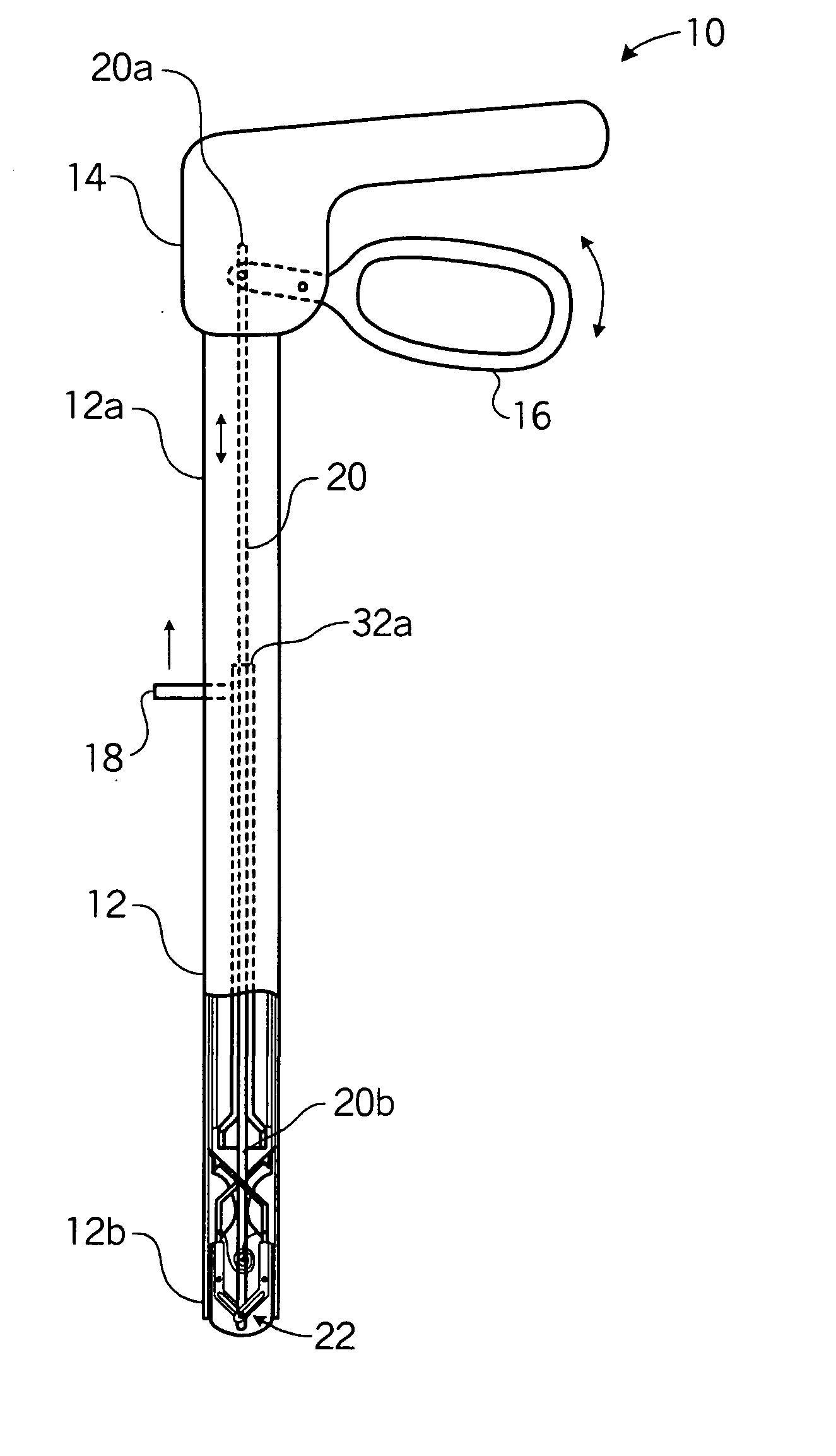
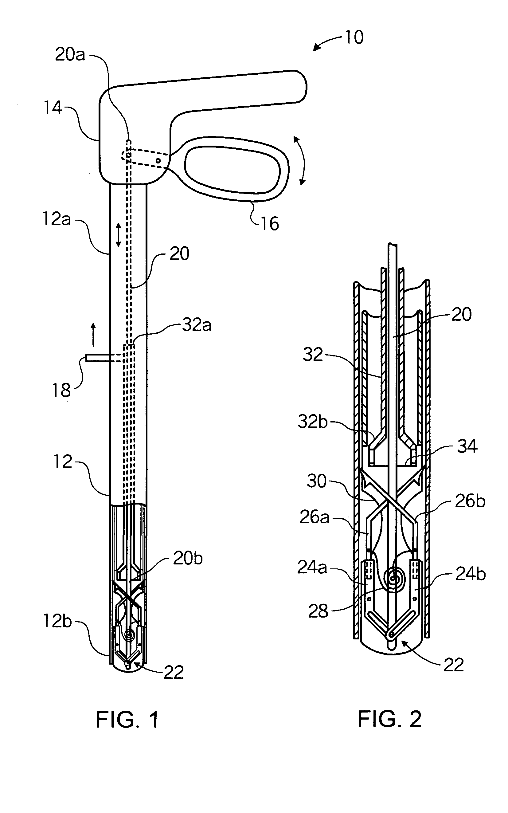
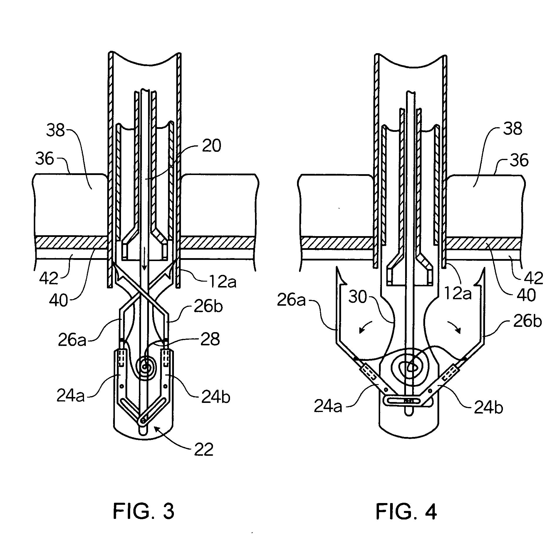
[0027] More specifically, the present invention is directed to a device that is operative to fashion a secure closure or suture of incisions or punctures, in organs, vasculature, and or tissues surrounding a cavity of the body. According to a preferred embodiment, the device comprises an elongate cannula or trocar or other hollow housing member having proximal and distal ends, the latter being configured to be inserted within...


PUM


Abstract

A laparoscopic fascial closure device (10) for fashioning a secure closure about a laparoscopic puncture site. The device comprises an elongate cannula (12) having proximal (12a) and distal (12b) ends. A needle suture complex (22) is selectively deployed from the distal end (12b) of the device that is operative to deploy a suture across the puncture site from within the body and draw the free ends of the suture outwardly from the body via the laparoscopic port site. The device (10) is configured to be utilized through a ten millimeter or larger laparoscopic port, and is operative to position the suture at the puncture site such that the suture extends in a diametrically-opposed configuration at least 1.0 cm or greater across opposed sides of the puncture site.

Description

RELATED APPLICATIONS AND CLAIMS OF PRIORITY [0001] This application claims priority to, and incorporates by reference in its entirety, co-pending U.S. patent application Ser. No. 10 / 086,318, filed Mar. 1, 2002, entitled “Laparoscopic Port Site Fascial Closure Device”.BACKGROUND OF THE INVENTION [0002] Laparoscopic, endoscopic, and thorascopic surgeries are well-known surgical techniques that advantageously reduces patient recovery time due to minimal tissue damage, which consequently permits the patient to return to normal activity in a shorter period of time. Generally, surgery relies upon the formation of one or more trocar puncture wounds through which are deployed surgical instruments and a rod-like telescope with a light source to enable the surgeon to view the organs and conduct the surgery. [0003] Notwithstanding the tremendous advantages afforded by such surgical techniques, there still presents substantial clinical problems. More specifically, the puncture wounds created-wi...

Claims


Application Information

Patent Timeline
Patent Type & Authority Applications(United States)
IPC IPC(8): A61B17/00A61B17/04A61B17/06A61B17/11
CPCA61B17/0057A61B17/0469A61B2017/00637A61B2017/06071A61B2017/0472A61B2017/0496A61B2017/00663
Inventor KERR, STEPHEN
Owner INTELLIMED SURGICAL SOLUTIONS
Why Eureka
Try Eureka