Device and method for intralumenal anastomosis

a technology of anastomosis and lumbar tube, which is applied in the field of surgery, can solve the problems of many non-operative therapies for morbid obesity that have been tried with virtually no permanent success, and failed to correct the condition

Active Publication Date: 2005-06-30
ETHICON ENDO SURGERY INC
View PDF26 Cites 150 Cited by
  • Summary
  • Abstract
  • Description
  • Claims
  • Application Information

AI Technical Summary

Benefits of technology

The invention is an introducer instrument that uses a special material called Shape Memory Alloy (SMA) to create an anastomosis device. This device can be placed within a patient's body through a minimally invasive laparoscopic puncture, and can even be done entirely through an endoscope. The SMA material has the ability to change its shape and can be shaped into a tightly coiled form to create a secure anastomosis. This technology allows for safer and more efficient medical procedures with minimal damage to the patient.

Problems solved by technology

The technical problem addressed in this patent text is the need for a better method of creating anastomosis for surgical procedures, such as gastric bypass, that can be performed with a minimum number of transcutaneous punctures and can potentially be performed on an out-patient basis. The current methods are time-consuming, technique-dependent, and may cause complications. The patent proposes a new approach to create an anastomosis device that can fully and accurately deploy at an anastomotic site between multiple lumens.

Method used


Image

  • Device and method for intralumenal anastomosis
  • Device and method for intralumenal anastomosis
  • Device and method for intralumenal anastomosis

Examples


Embodiment Construction

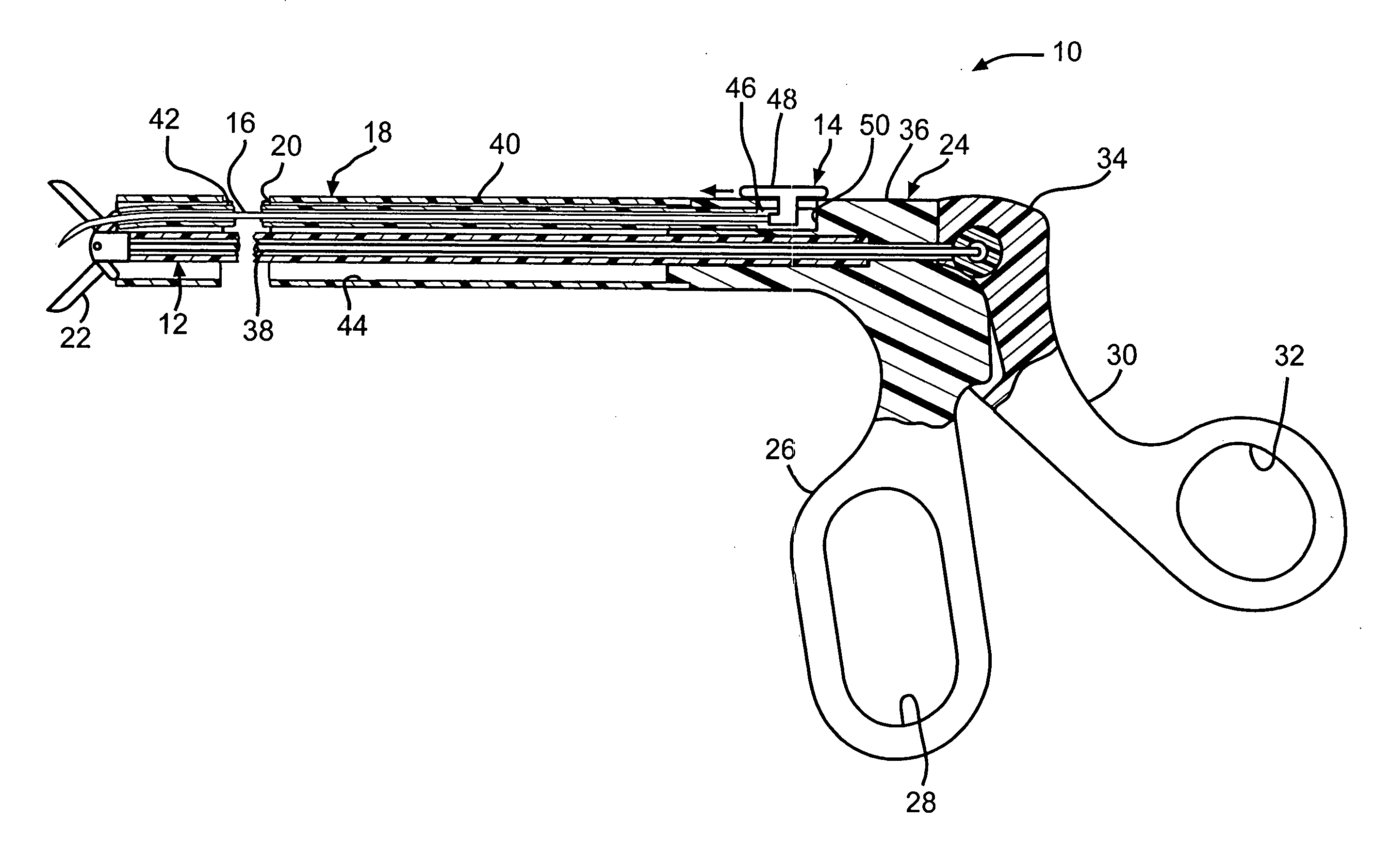
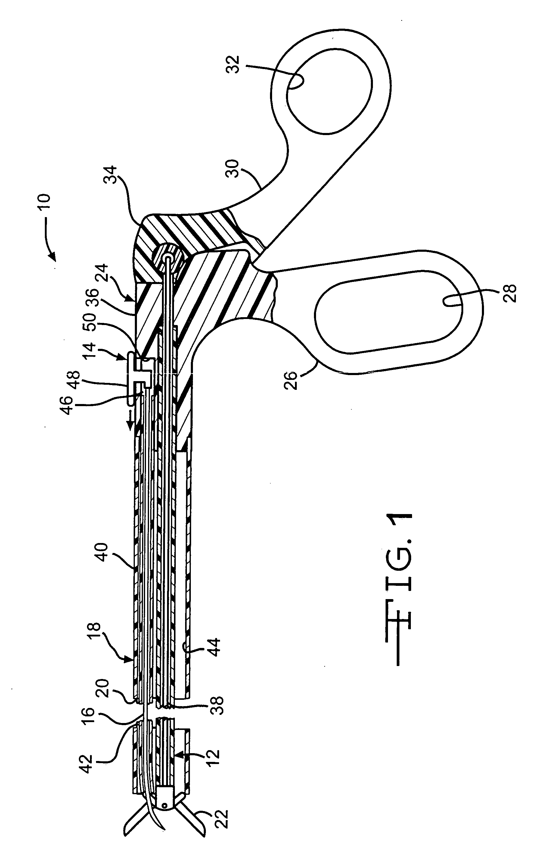
[0029] Turning to the drawings, wherein like numerals denote like components throughout the several views, in FIG. 1, an anastomosis instrument 10 advantageously incorporates both a grasping mechanism 12 as well as an extrusion mechanism 14 for dispensing a straightened anastomosis device (“clamp”) 16 for piercing two tissue walls (not shown in FIG. 1) of adjacent lumens. The anastomosis device 16 is formed of a Shape Memory Alloy (SMA) that was annealed into a flattened coil and then straightened. After dispensing, the anastomosis device 16 thereafter responds to no longer being constrained by the anastomosis instrument 10 and by being warmed by body heat. Thereafter, tightened coils are formed to provide an accurate and consistent anastomosis with a patent lumen in an endoscopic setting, perhaps obviating the need for laparoscopic punctures.

[0030] In the version of the anastomosis instrument 10 depicted in FIG. 1, the grasping mechanism 12 is similar to generally known graspers o...


PUM


Abstract

An anastomosis device formed from a Shape Memory Alloy (SME) wire that is annealed into a tight coil (e.g., flat coil, helical coil, cylindrical coil) that is subsequently substantially straightened (e.g., straight, longitudinally stretched spring-like shape) for being constrained within an elongate member of an anastomosis introducer instrument. After positioning and holding two tissue walls of adjacent lumens into apposition with a distally presented grasper of the instrument, a piercing tip of the anastomosis device is dispensed and inserted through the tissue walls before being allowed to relax to its tight coil shape, thereby forming the anastomosis attachment. The anastomosis device is released from the anastomosis introducer instrument, such as by fully dispensing its proximal end. Thereby, efficient single lumen anastomosis of gastrointestinal, bilatory, or other vessels may be achieved with a minimum of laparoscopic punctures, or perhaps performed fully as an endoscopic procedure.

Description


Claims


Application Information

Patent Timeline
Owner ETHICON ENDO SURGERY INC
Why Eureka
Try Eureka