Clinical tool for structure localization

a technology of structure localization and clinical tools, applied in the field of clinical tools for structure localization, data collection and processing methods, can solve the problems of considerable skill and intuition, the most time-consuming portion of the procedure, and the most difficult to achiev

Inactive Publication Date: 2005-07-07
MEDTRONIC INC
View PDF2 Cites 30 Cited by
  • Summary
  • Abstract
  • Description
  • Claims
  • Application Information

AI Technical Summary

Problems solved by technology

This maneuvering is often the most time-consuming portion of the procedure as it can require considerable skill and intuition, particularly when the intra-operative images are two-dimensional images.

Method used


Image

  • Clinical tool for structure localization
  • Clinical tool for structure localization
  • Clinical tool for structure localization

Examples


Embodiment Construction

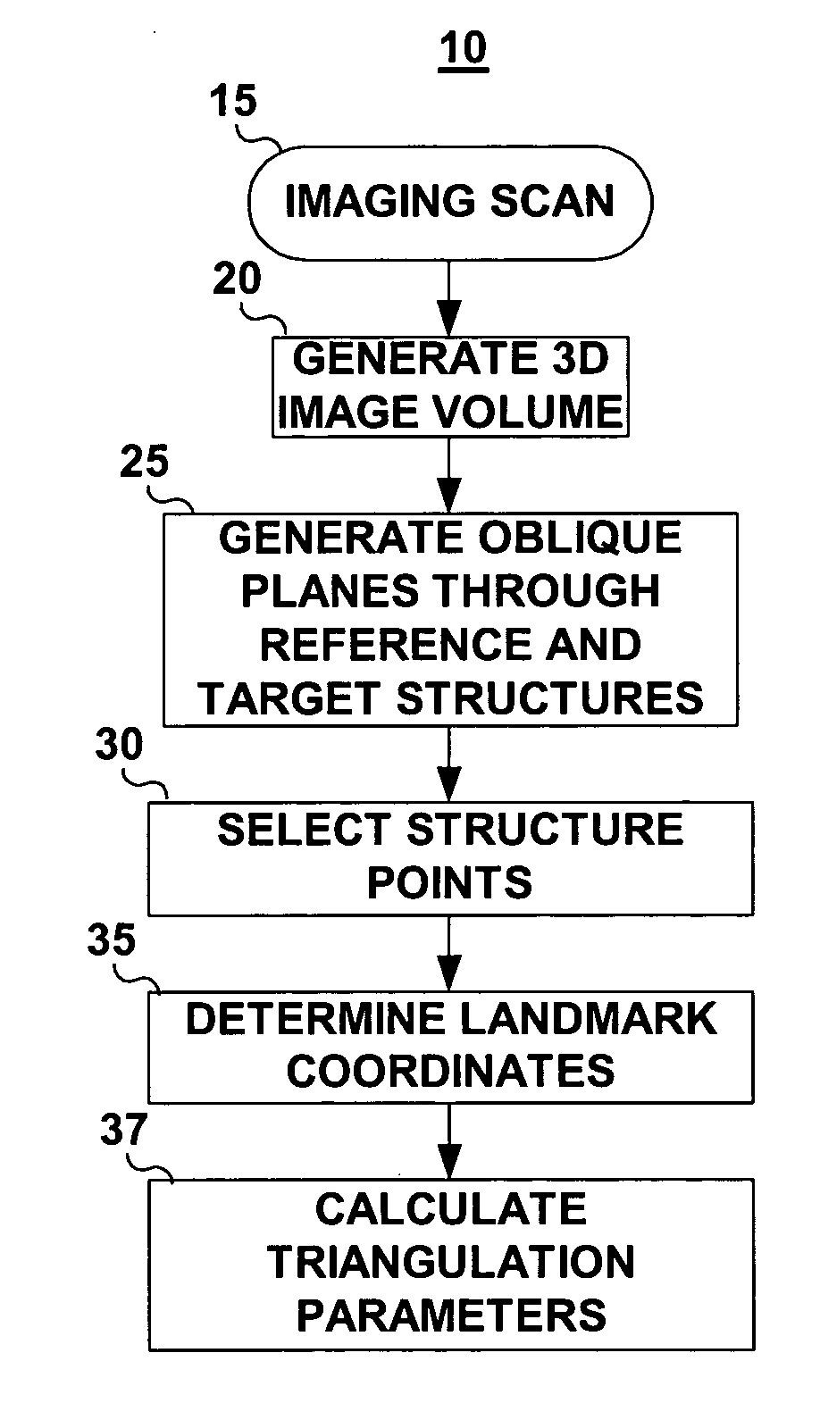
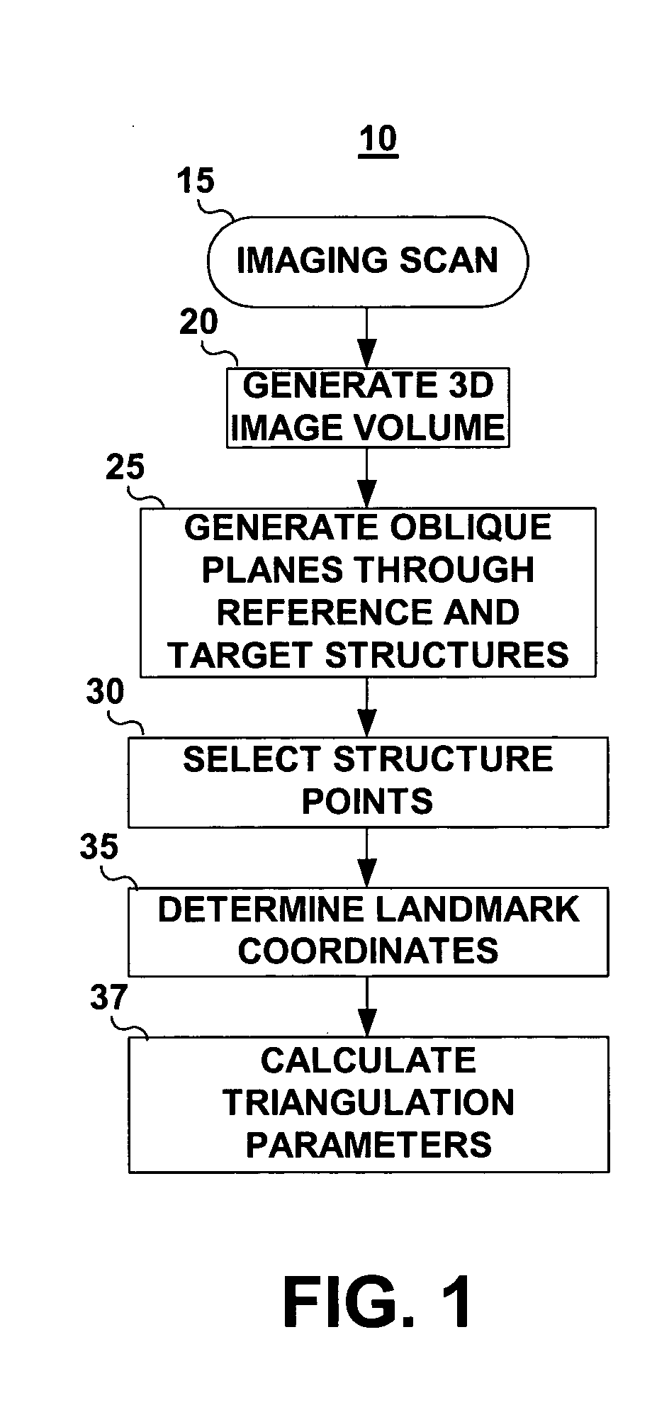
[0019] The invention provides clinicians with a useful tool for localizing internal structures particularly during minimally invasive procedures during which the clinician does not have direct line of sight view of a targeted structure. The targeted structure may be an anatomical structure, a pathological feature, or an implanted device. During a minimally invasive procedure, the clinician is typically guiding an instrument or medical device toward the targeted structure by viewing the position of the instrument in a 2D or 3D image obtained intra-operatively. Such procedures can be time-consuming and require considerable skill and intuition. The invention provides a method for plotting an estimated location of a targeted structure on intra-operative medical images to aid the clinician in localizing the targeted structure. The plotted location is estimated based on previously collected triangulation parameter data relating the targeted structure location to selected reference structu...


PUM


Abstract

A method for localizing an internal body structure during an image-guided procedure that includes obtaining a three dimensional image of a body portion containing a targeted structure and selected reference structures; determining a set of landmark point coordinates for defining the location of each of the targeted and reference structures; computing triangulation parameters relating the location of the landmark points for the targeted structure and the reference structures; and, during an image-guided procedure, using the computed triangulation parameters to plot an estimated location of the targeted structure on an intra-operative image in which the reference structures have been identified.

Description

CLAIM OF PRIORITY [0001] This application claims priority from provisional Application Ser. No. 60 / 520,358, filed Nov. 13, 2003, entitled “CLINICAL TOOL FOR STRUCTURE LOCATION.”FIELD OF THE INVENTION [0002] The present invention relates to methods for guiding medical interventions using anatomical or pathological landmarks and more particularly to a data collection and processing method that facilitates image-guided surgery. BACKGROUND OF THE INVENTION [0003] Minimally invasive techniques are being developed for a variety of surgical procedures. During such procedures, the clinician typically relies on intra-operative imaging to guide instrumentation to a targeted therapy or diagnostic site. This maneuvering is often the most time-consuming portion of the procedure as it can require considerable skill and intuition, particularly when the intra-operative images are two-dimensional images. With the increasing use of minimally invasive techniques, clinical tools are needed that aid a c...

Claims


Application Information

Patent Timeline
Patent Type & Authority Applications(United States)
IPC IPC(8): A61B5/05A61B5/06
CPCA61B5/7285A61B5/06A61B5/065
Inventor LAHM, RYAN P.BAXTER, WALTON W. IIIMORISSETTE, JOSEELASKE, TIMOTHY G.
Owner MEDTRONIC INC
Why Eureka
Try Eureka