

Computerized scheme for distinction between benign and malignant nodules in thoracic low-dose CT
a computerized scheme and low-dose ct technology, applied in the field of automatic detection of structures and abnormalities in medical images, can solve the problems of difficulty for radiologists to distinguish between benign and malignant nodules on ldct, and the method of suzuki et al. is not capable of providing a continuous scor
- Summary
- Abstract
- Description
- Claims
- Application Information
AI Technical Summary
Problems solved by technology
Method used
Image

Examples
Embodiment Construction
[0085] In describing preferred embodiments of the present invention illustrated in the drawings, specific terminology is employed for the sake of clarity. However, the invention is not intended to be limited to the specific terminology so selected, and it is to be understood that each specific element includes all technical equivalents that operate in a similar manner to accomplish a similar purpose. Moreover, features and procedures whose implementations are well known to those skilled in the art, such as initiation and testing of loop variables in computer programming loops, are omitted for brevity.
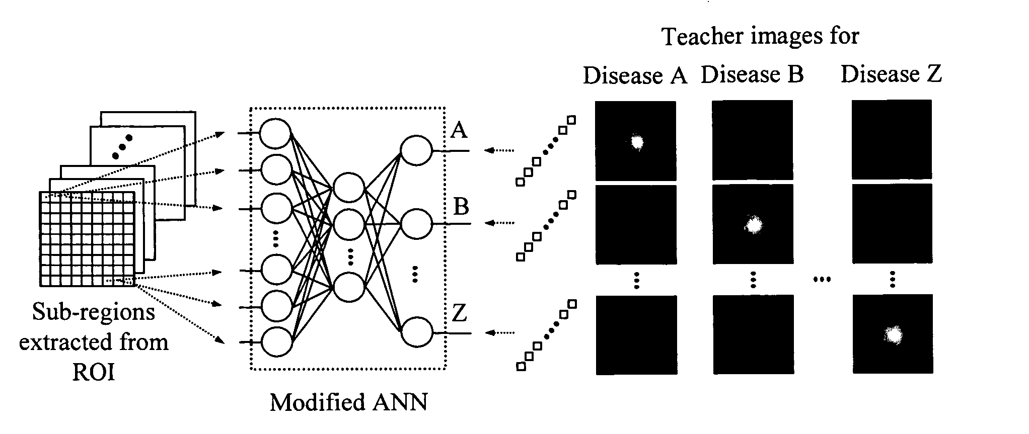
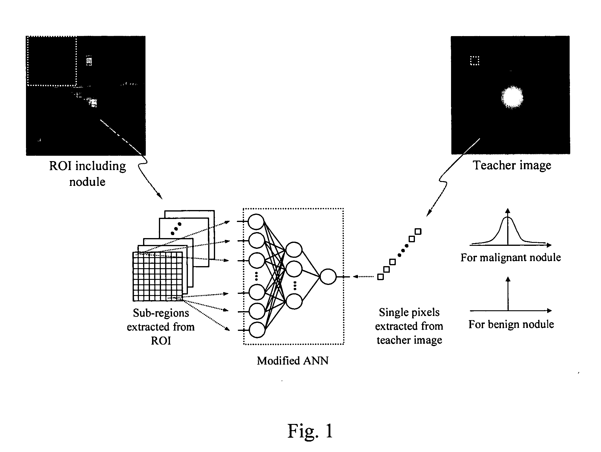
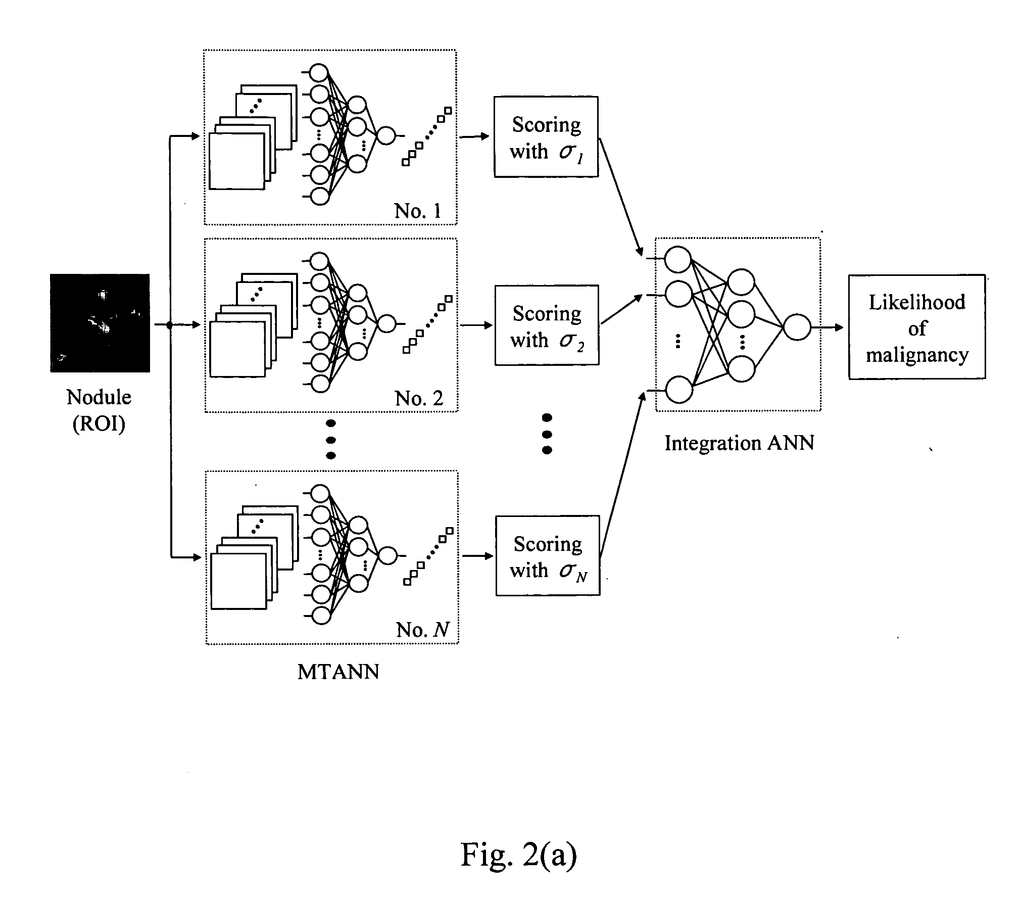
[0086] The present invention provides various image-processing and pattern recognition techniques in arrangements that may be called a massive training artificial neural networks (MTANNs) and their extension, Multi-MTANNs.
[0087] For the purposes of this description an image is defined to be a representation of a physical scene, in which the image has been generated by some imaging tec...
PUM
 Abstract
Description
Claims
Application Information