System and method for three-dimensional space management and visualization of ultrasound data ("SonoDEX")

a three-dimensional space and data visualization technology, applied in the field of interactive display and manipulation of a three-dimensional space, can solve the problem that users do not gain a 3d sense of where the ultrasound slice fits in vis-a-vis the patien

Inactive Publication Date: 2006-01-26
BRACCO IMAGINIG SPA
View PDF13 Cites 183 Cited by
  • Summary
  • Abstract
  • Description
  • Claims
  • Application Information

AI Technical Summary

Benefits of technology

[0012] A system and method for the imaging management of a 3D space where various substantially real-time scan images have been, or are being, acquired are presented. In exemplary embodiments of the present invention, a user can visualize images of a portion of a body or object obtained from a substantially real-time scanner not just as 2D images, but as positionally and orientationally identified slices within the relevant 3D space. In exemplary embodiments of the present invention, a user can convert such slices into volumes as desired, and can process the images or volumes using known image processing and / or volume rendering techniques. Alternatively, a user can acquire ultrasound images in 3D using the techniques of UltraSonar or 4D Ultrasound. In exemplary embodiments of the present invention, a user can manage various substantially real-time images that have been obtained, either as slices or volumes, and can control their visualization, processing and display, as well as their registration and fusion with other images, volumes or virtual objects obtained or derived from prior scans of the area or object of interest using various modalities.

Problems solved by technology

Nonetheless, while the individual slices comprising such a volume are loosely registered to each other (loosely because the subject's body is moving throughout the acquisition, and thus the body does not have a fixed spatial relationship to the probe during the acquisition) they are not registered in any sense to the 3D patient space.
Moreover, even if such a volume is acquired and displayed, the physical interfaces provided to manipulate these volumes are not themselves three-dimensional, generally being nothing more than a standard computer keyboard and mouse (or the equivalent, such as a trackball).
Accordingly, using such tools to effect 3D operations necessitates awkward mappings of 3D manipulations onto essentially 2D devices.
The necessity of such awkward mappings may be one of the reasons why 3D visualization has not gained the acceptance in the medical community that it may be due.
However, because in such systems the display of ultrasound is restricted to the plane of acquisition, the pre-scan data is provided as 2D slices that match the plane of the ultrasound slice, and the ultrasound and corresponding pre-operative scan cut are simply placed side-by-side for comparison, a user does not gain a 3D sense of where the ultrasound slice fits in vis-a-vis the patient space as a whole.

Method used


Image

  • System and method for three-dimensional space management and visualization of ultrasound data ("SonoDEX")
  • System and method for three-dimensional space management and visualization of ultrasound data ("SonoDEX")
  • System and method for three-dimensional space management and visualization of ultrasound data ("SonoDEX")

Examples


Embodiment Construction

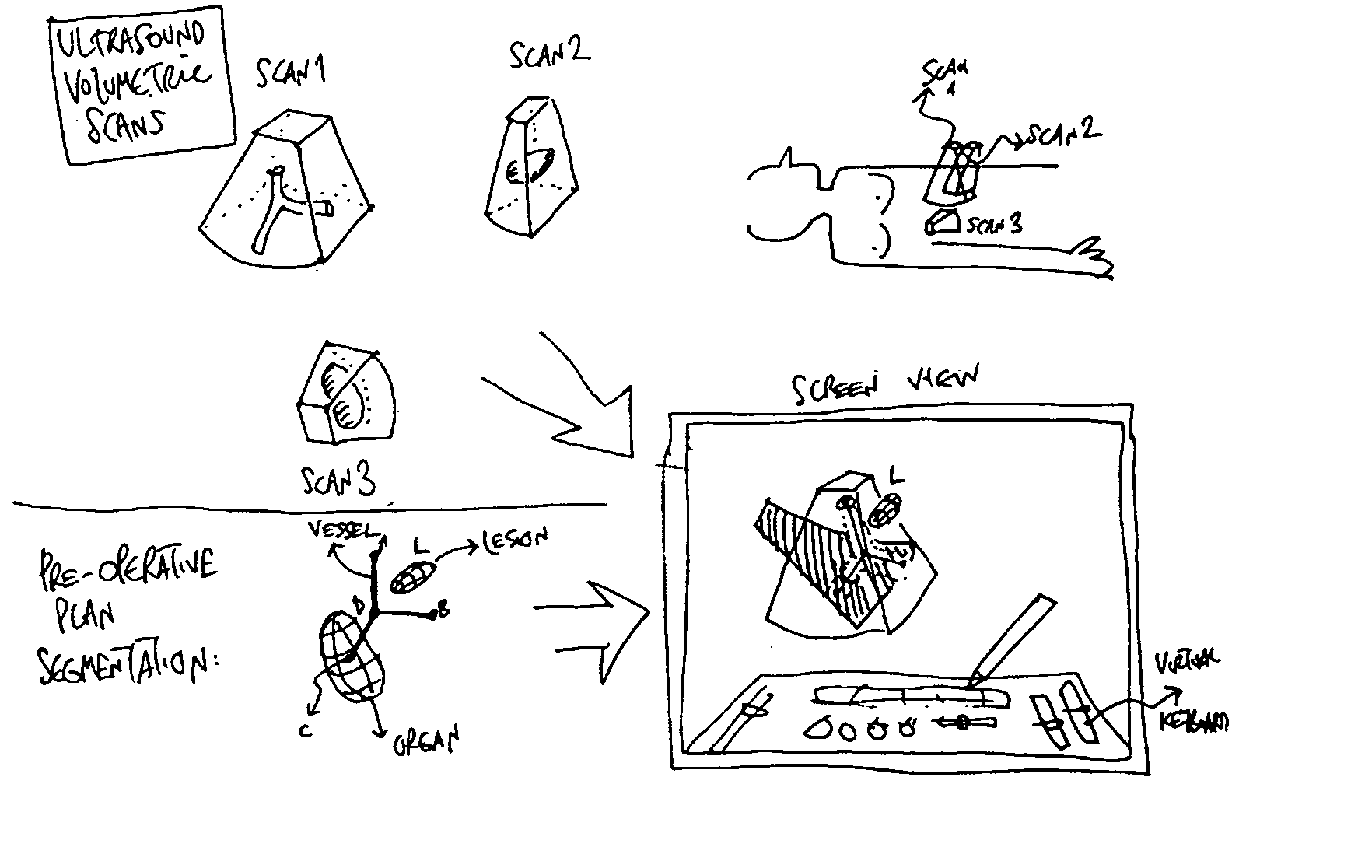
[0034] This present invention is directed to a system and method for the management of a 3D space where substantially real-time images have been, or are being, acquired. For purposes of illustration, exemplary embodiments of the invention will be described with reference to ultrasound images, it being understood that any equivalent substantially real-time imaging modality can be used.

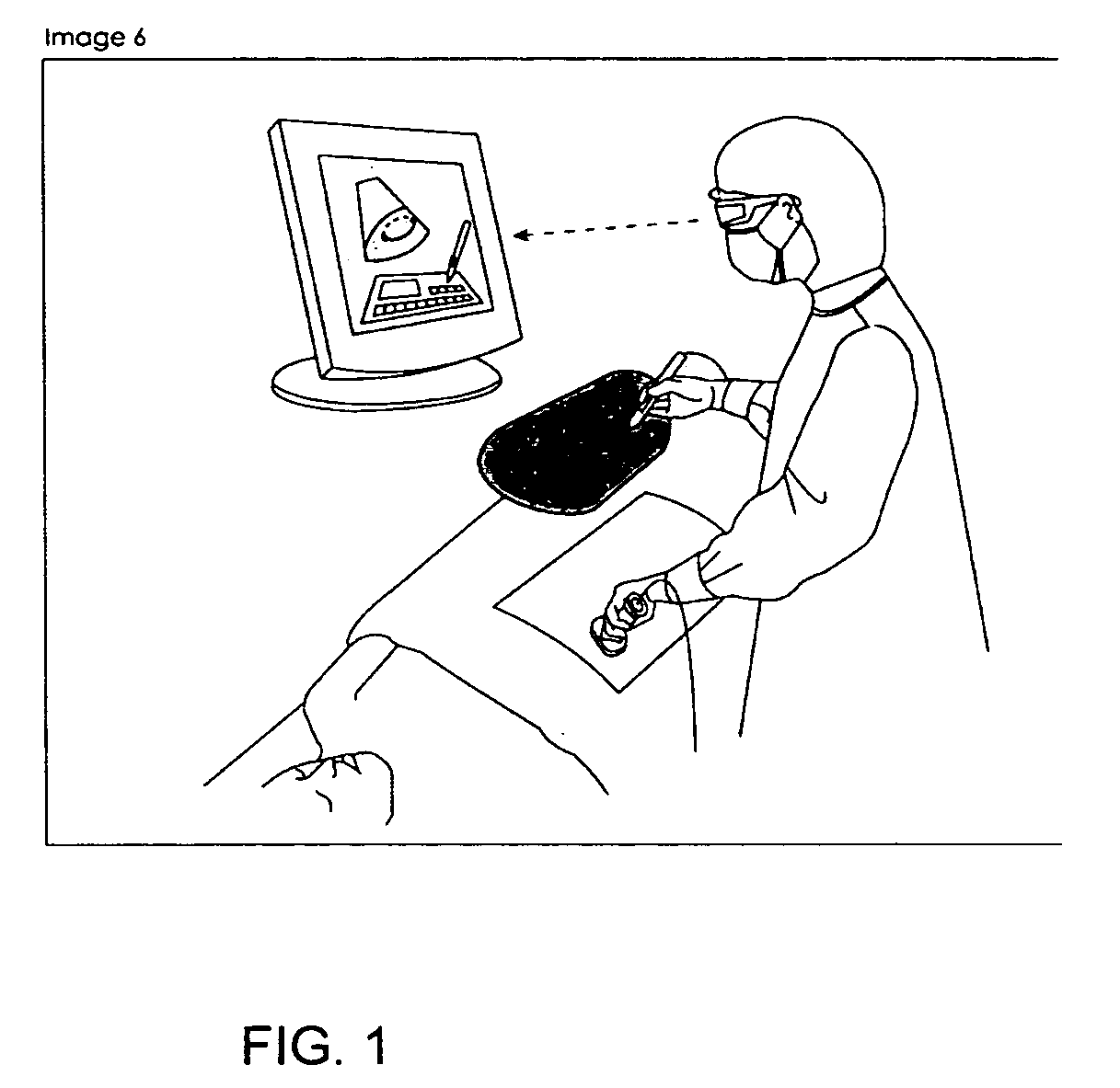
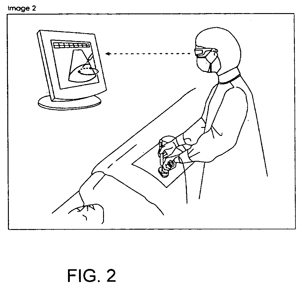
[0035] In exemplary embodiments of the present invention a clinician can visualize images obtained from an ultrasound scanner not just as 2D images but as 2D slices within a particular 3D space (or alternatively as volumes within such 3D space), each acquired at a known time, and can convert such 2D slices into volumes whenever needed. In exemplary embodiments of the present invention, the method allows a user to manage the different images obtained (either as slices or volumes), and to manipulate them as well as control various display parameters, for example, their visualization (including stereoscop...


PUM


Abstract

A system and method for the imaging management of a 3D space where various substantially real-time scan images have been acquired is presented. In exemplary embodiments according to the present invention, a user can visualize images of a portion of a body or object obtained from a substantially real-time scanner not just as 2D images, but as positionally and orientationally located slices within a particular 3D space. In such exemplary embodiments a user can convert such slices into volumes whenever needed, and can process the images or volumes using known image processing and/or volume rendering techniques. Alternatively, a user can acquire ultrasound images in 3D using the techniques of UltraSonar or 4D Ultrasound. In exemplary embodiments according to the present invention, a user can manage various substantially real-time images obtained, either as slices or volumes, and can control their visualization, processing and display, as well as their registration and fusion with other images, volumes and virtual objects obtained or derived from prior scans of the body or object of interest using various modalities.

Description

CROSS REFERENCE TO RELATED APPLICATIONS [0001] This application claims the benefit of the following U.S. Provisional Patent Applications: (i) Ser. No. 60 / 585,214, entitled “SYSTEM AND METHOD FOR SCANNING AND IMAGING MANAGEMENT WITHIN A 3D SPACE (“SonoDEX”)”, filed on Jul. 1, 2004; (ii) Ser. No. 60 / 585,462, entitled “SYSTEM AND METHOD FOR A VIRTUAL INTERFACE FOR ULTRASOUND SCANNERS (“Virtual Interface”)”, filed on Jul. 1, 2004; and (iii) Ser. No. 60 / 660,858, entitled “SONODEX: 3D SPACE MANAGEMENT AND VISUALIZATION OF ULTRASOUND DATA”, filed on Mar. 11, 2005. [0002] The following related United States patent applications, under common assignment herewith, are also fully incorporated herein by this reference: Ser. No. 10 / 469,294 (hereinafter “A Display Apparatus”), filed on Aug. 29, 2003; Ser. No. 10 / 725,773 (hereinafter “Zoom Slider”), Ser. No. 10 / 727,344 (hereinafter “Zoom Context”), and Ser. No. 10 / 725,772 (hereinafter “3D Matching”), each filed on Dec. 1, 2003; Ser. No. 10 / 744,869 ...

Claims


Application Information

Patent Timeline
Patent Type & Authority Applications(United States)
IPC IPC(8): A61B8/00
CPCA61B5/201G06T2210/41A61B8/0833A61B8/0841A61B8/0891A61B8/14A61B8/4245A61B8/4416A61B8/481A61B8/483G01S7/52034G01S7/52063G01S7/52068G01S7/5208G02B27/017G02B2027/0134G02B2027/014G06T7/0038G06T19/00G06T2207/30004A61B8/06A61B8/4254G06T7/38
Inventor SERRA, LUISCHOON, CHUA BENG
Owner BRACCO IMAGINIG SPA
Why Eureka
Try Eureka