

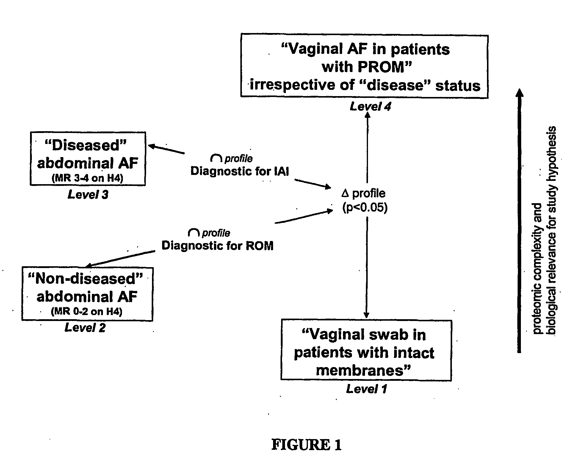
Non-invasive assessment of intra-amniotic environment
a non-invasive, amniotic environment technology, applied in the direction of material analysis, biological material analysis, instruments, etc., can solve the problems of reducing the risk of chorioamnionitis without increasing the overall cesarean delivery rate, increasing the probability of appropriate treatment, and disappointing results
- Summary
- Abstract
- Description
- Claims
- Application Information
AI Technical Summary
Benefits of technology
Problems solved by technology
Method used
Image

Examples
example 1
Discovery of Biomarkers by Comparing Vaginal Samples from Patients with Normal and Abnormal Clinical Status
[0100] Biomarkers according to the present invention were identified by comparing mass spectra of samples derived from vaginal fluid from two groups of pregnant subjects, subjects with an abnormal clinical status and normal subjects. The subjects were diagnosed according to standard clinical criteria. The two different pools of samples enable a differential analysis. Alternatively, surrogate samples were produced in vitro. For example, to confirm intra-amniotic bleeding the amniotic fluid profile in patients with an amniotic fluid red blood cell count over 5000 cells / mm3 was intersected with a diluted red blood cell lysate obtained from umbilical cord blood (fetal origin).
[0101] The two sample pools were used in a wide range of dilutions to test various biochip surfaces, produced by Ciphergen Biosystems, Fremont, Calif., for optimal discriminatory performance, including rever...
example 2
Discovery of Biomarkers by Comparing Vaginal Samples from Patients with Normal and Abnormal Clinical Status with Contemporaneously-Obtained Samples of Amniotic Fluid Via Amniocentesis
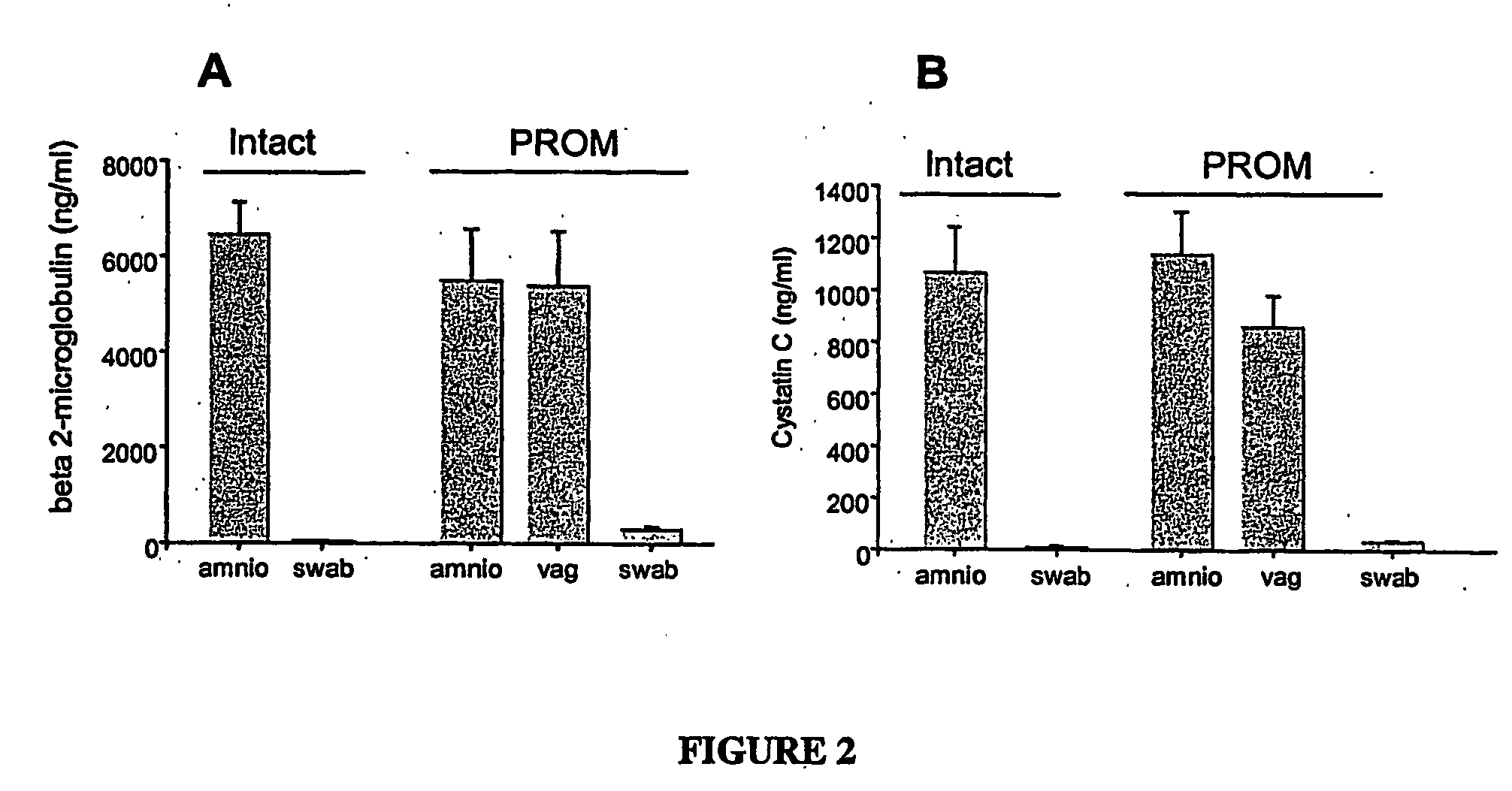
[0113] Samples obtained by a swab of the vagina of patients with intact or ruptured membranes and samples of amniotic fluid obtained contemporaneously by amniocentesis from the same patient were compared using specific ELISA assays. The results are shown in FIG. 2. When a swab saturated with amniotic fluid is immersed in 0.5 ml PBS the concentration in swab fluid of beta-2 microglobulin is 10% of the concentration in the pure fluid. This demonstrates that in women with ruptured membranes the vaginal environment contains biomarkers of amniotic cavity origin, such as beta-2-microglobulin and cystatin C, in a similar concentration to that found in the amniotic fluid obtained by amniocentesis. These biomarkers are absent in the vagina of women with intact membranes. Results with the SELDI platform showed t...
example 3
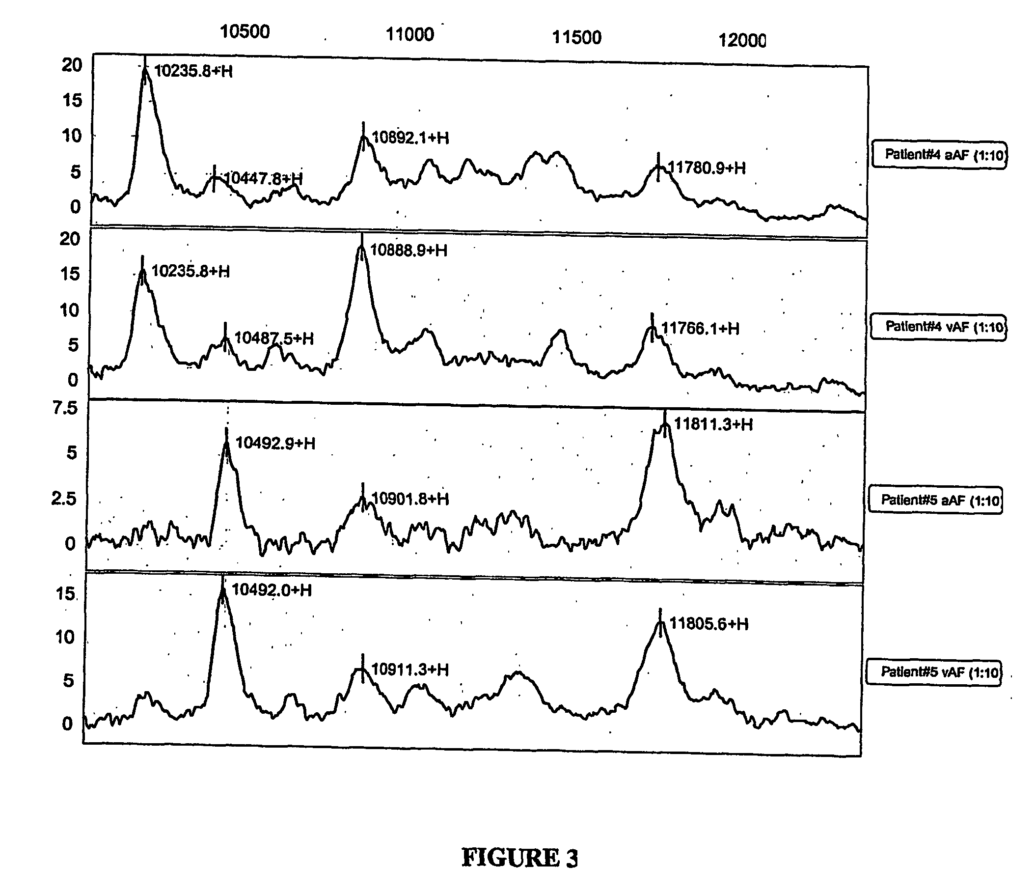
Serial Monitoring of Intra-Amniotic Inflammation
[0115] Serial samples of vaginal fluid from pregnant women were obtained and profiled by SELDI. This serial sampling successfully identified the appearance of inflammatory biomarkers indicative of incipient intra-amniotic inflammation before there was any clinical evidence. A normal profile obtained from the patient's vagina before rupture was subtracted to allow identification of the amniotic fluid peaks characteristics of inflammation, such as beta-2-microglobulin, cystatin C, alpha-fetoprotein. Even though the fluid became sparse (anhydramnios) as time passed, and the vaginal fluid became more viscous, profiling of samples by SELDI was possible. In this case, the sample was obtained by washing the syringe used for sampling with PBS.
[0116]FIG. 4 shows a protein profile obtained from vaginal fluid of a patient monitored serially. On the day of admission, her normal profile was dominated by a high beta 2-microglobulin peak (upper tra...
PUM
 Abstract
Description
Claims
Application Information