Method and apparatus for generating a 2D image having pixels corresponding to voxels of a 3D image

a 3d image and image technology, applied in the field of medical imaging, can solve the problem of difficult segmentation of coronary arteries

Inactive Publication Date: 2006-11-09
SIEMENS MEDICAL SOLUTIONS USA INC
View PDF7 Cites 98 Cited by
  • Summary
  • Abstract
  • Description
  • Claims
  • Application Information

AI Technical Summary

Problems solved by technology

Coronary arteries are typically difficult to segment because of their size and proximity to the surface of the heart and blood pool.

Method used


Image

  • Method and apparatus for generating a 2D image having pixels corresponding to voxels of a 3D image
  • Method and apparatus for generating a 2D image having pixels corresponding to voxels of a 3D image
  • Method and apparatus for generating a 2D image having pixels corresponding to voxels of a 3D image

Examples


Embodiment Construction

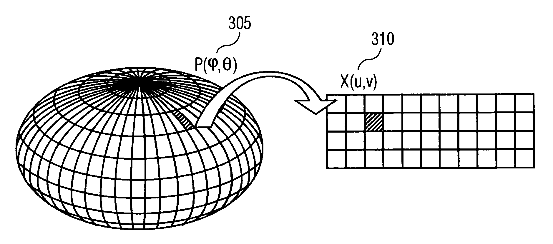
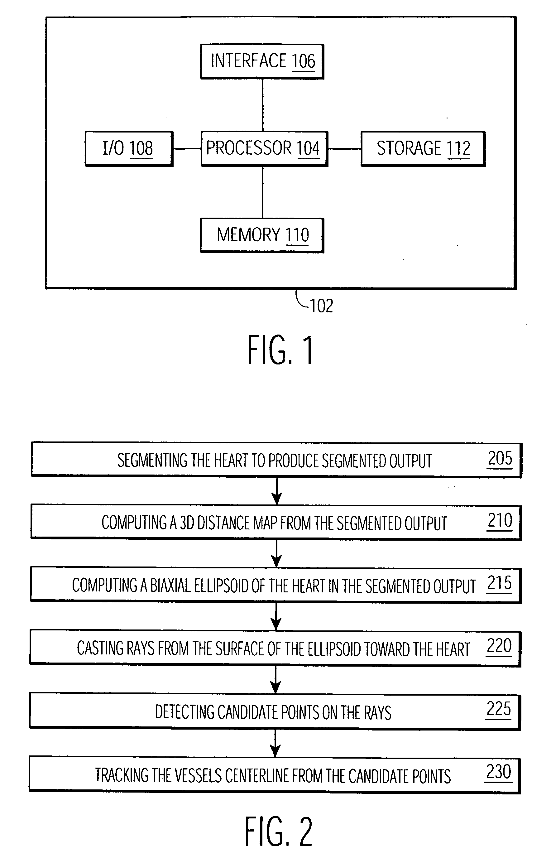
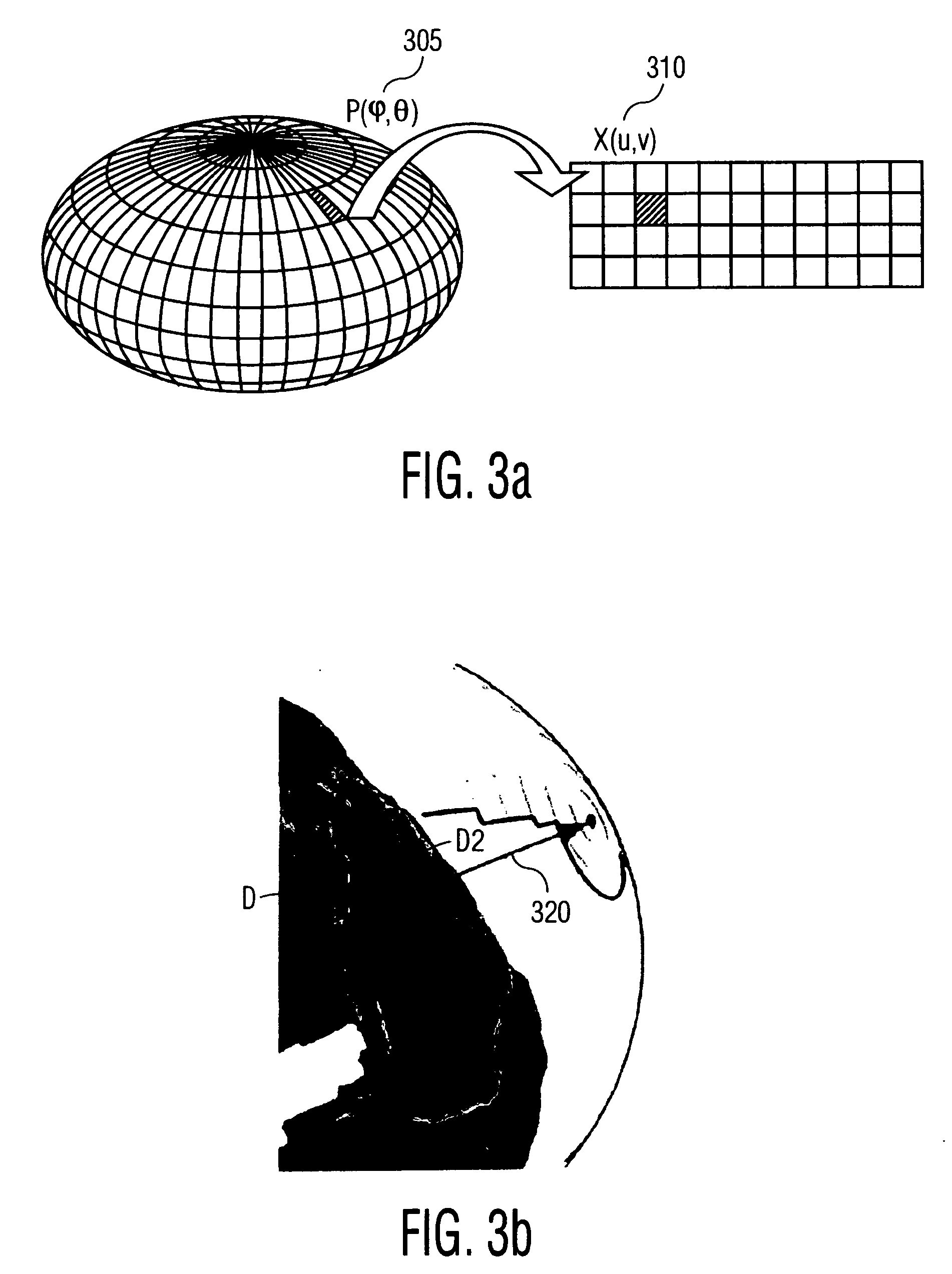
[0021] The following description describes the present invention in terms of the processing steps required to implement an embodiment of the invention. These steps may be performed by an appropriately programmed computer, the configuration of which is well known in the art. An appropriate computer may be implemented, for example, using well known computer processors, memory units, storage devices, computer software, and other components. A high level block diagram of such a computer is shown in FIG. 1. Computer 102 contains a processor 104 which controls the overall operation of computer 102 by executing computer program instructions which define such operation. The computer program instructions may be stored in a storage device 112 (e.g., magnetic disk) and loaded into memory 110 when execution of the computer program instructions is desired. Computer 102 also includes one or more interfaces 106 for communicating with other devices (e.g., locally or via a network). Computer 102 als...


PUM


Abstract

Disclosed is a method and apparatus for generating a two dimensional (2D) image of a structure (e.g., an organ) that has at least one pixel corresponding to at least one voxel of a three dimensional (3D) image of the structure. First, the surface of the structure in the 3D image is modeled by a geometrical volume such as an ellipsoid. Next, normal maximum intensity projection (MIP) rays are cast (i.e., projected) for voxels of the geometrical volume. The 2D image is then generated using the rays. The 2D image has at least one pixel that corresponds to at least one voxel of the 3D image.

Description

[0001] This application claims the benefit of U.S. Provisional Application No. 60 / 670,889 filed Apr. 13, 2005, which is incorporated herein by reference.BACKGROUND OF THE INVENTION [0002] The present invention relates generally to medical imaging, and more particularly to generating a 2 dimensional (2D) image having pixels that correlate to voxels of a three dimensional (3D) image. [0003] Cardiovascular diseases, such as heart disease and stroke, are one of the major causes of death in the U.S. To diagnose a cardiovascular disease, medical personnel may perform a Computed Tomography (CT) scan on a patient. A CT scan uses x-ray equipment to obtain image data from different angles around the human body and then processes the data to show a cross-section of the body tissues and organs. The image can then be analyzed by methods using morphological operators to highlight specific areas so that radiologists (or other medical personnel) can more easily diagnose problems such as cardiovascu...

Claims


Application Information

Patent Timeline
Patent Type & Authority Applications(United States)
IPC IPC(8): G06K9/00G06T15/08
CPCG06T15/08G06T11/008
Inventor FLORIN, CHARLESMOREAU-GOBARD, ROMAINWILLIAMS, JAMES P.
Owner SIEMENS MEDICAL SOLUTIONS USA INC
Why Eureka
Try Eureka