Contrast-enhanced cone beam X-ray imaging, evaluation, monitoring and treatment delivery

a cone beam and computed tomography technology, applied in the field of contrast-enhanced cone beam computed tomography, can solve the problems of low specificity of gd-mri, limited prediction value and specificity of x-ray mammography, and many false positives

Active Publication Date: 2007-11-22
VARIAN MEDICAL SYSTEMS +2
View PDF5 Cites 102 Cited by
  • Summary
  • Abstract
  • Description
  • Claims
  • Application Information

AI Technical Summary

Problems solved by technology

However, the predictive value and specificity of X-ray mammography are limited due to projecting a three-dimensional anatomy into a two-dimensional image and due to poor contrast detectability.
Ten further volume doubling times will lead to a tumor weighing about 1 kg with a size of 8-10 cm in diameter, which may be lethal.
However Gd-MRI suffers from low specificity with many false positive findings, where a benign tissue is incorrectly identified as malignant.
This is a problem shared with conventional compression X-ray projection mammography where about 75 percent of the biopsies of mammogram-identified suspicious regions turn out to be benign.
One problem with conventional cone beam CT is that the cancer lesions in the breast have about the same X-ray absorption coefficient and contrast level in CT slice images as the benign glandular tissue, which makes it difficult to differentiate the malignant lesions from benign structures, especially when the malignant lesions are adjacent to or combined with the glandular tissue.
This is particularly a problem in younger women with denser breasts that are comprised heavily of glandular tissue.

Method used


Image

  • Contrast-enhanced cone beam X-ray imaging, evaluation, monitoring and treatment delivery
  • Contrast-enhanced cone beam X-ray imaging, evaluation, monitoring and treatment delivery
  • Contrast-enhanced cone beam X-ray imaging, evaluation, monitoring and treatment delivery

Examples


Embodiment Construction

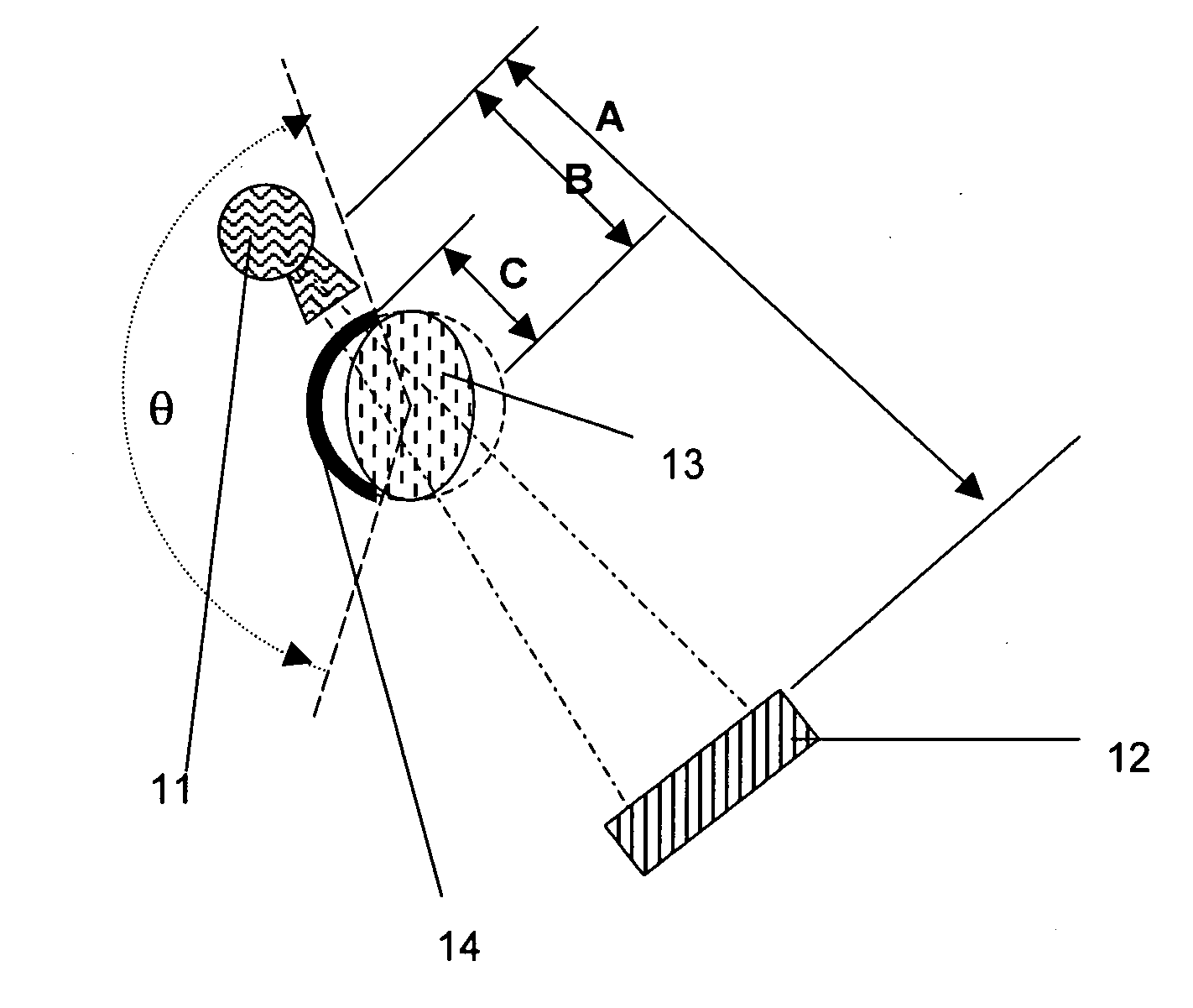
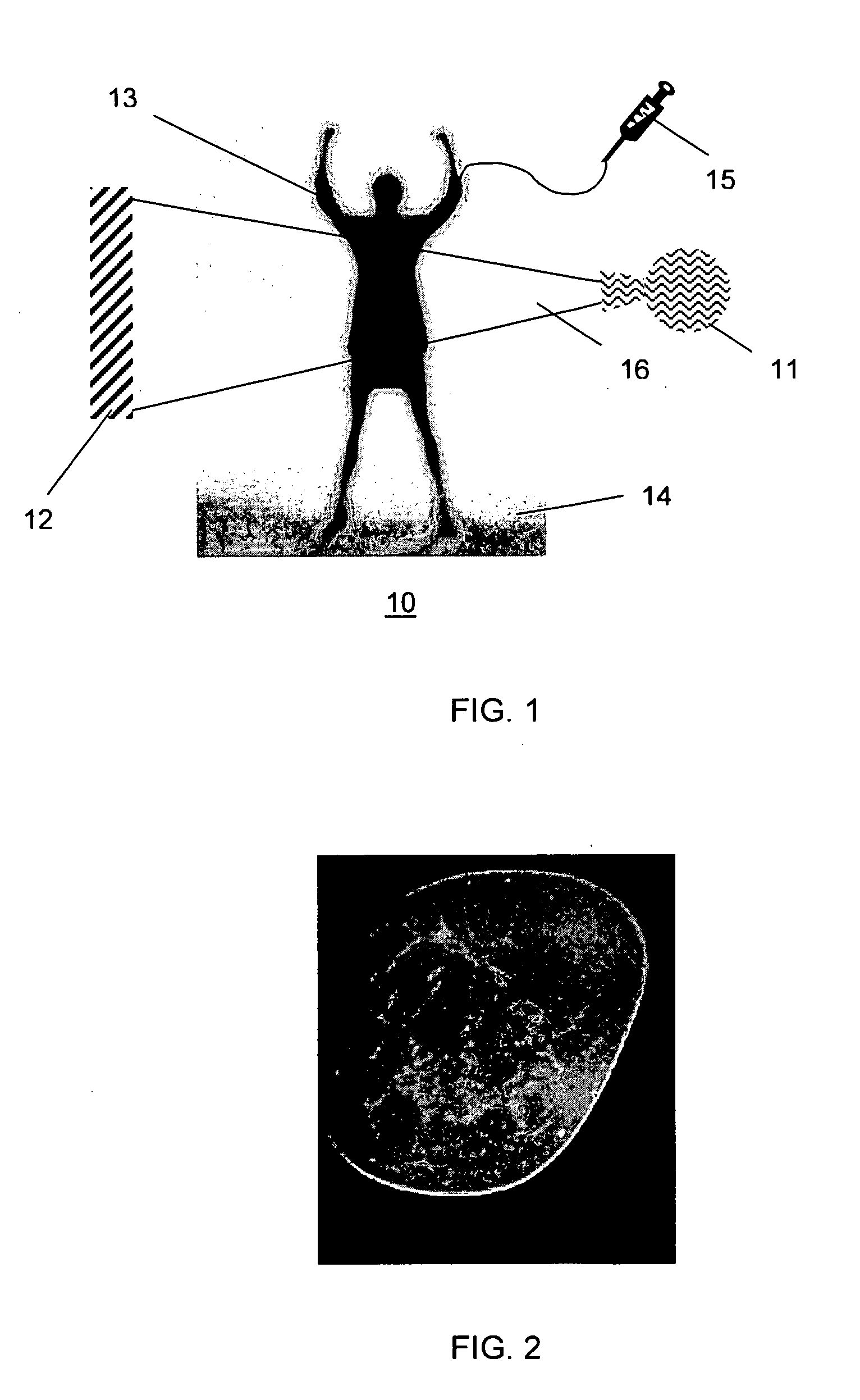
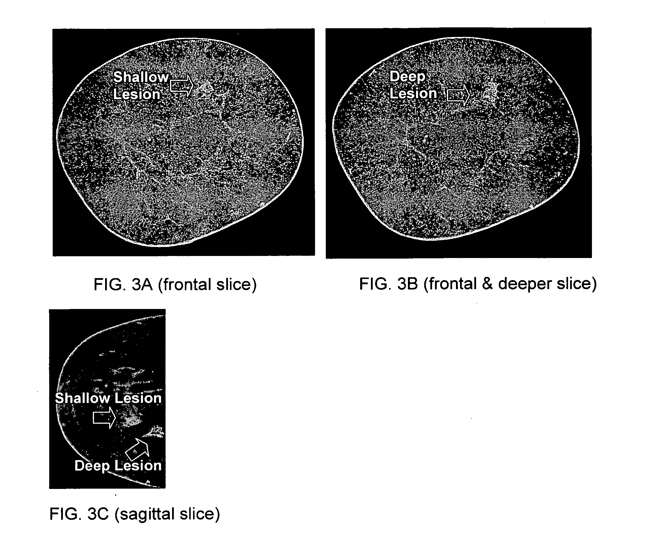
[0029] Various embodiments of the present invention are described hereinafter with reference to the figures. It should be noted that some figures are schematic and the figures are only intended to facilitate the description of specific embodiments of the invention. They are not intended as an exhaustive description of the invention or as a limitation on the scope of the invention. In addition, an aspect described in conjunction with a particular embodiment of the present invention is not necessarily limited to that embodiment and can be practiced in any other embodiments of the present invention. For instance, in the following description, the present invention is described with embodiments of cone beam computed tomography. It will be appreciated that the claimed invention may also be used with other X-ray imaging systems such as cone beam digital tomosynthesis, and inverse-geometry volumetric computed tomography (IGVCT) which is not cone beam based. Further, in the following descri...


PUM


Abstract

A method of imaging a patient's uncompressed region of interest using X-ray cone beam computed tomography or cone beam digital tomography comprises the step of introducing an effective amount of a contrast agent to the uncompressed region of interest. A system for imaging a patient's uncompressed region of interest using cone beam computed tomography (CBCT) or cone beam digital tomography (CBDT) comprises an X-ray source transmitting an X-ray to the uncompressed region of interest, an image acquisition system acquiring a plurality of two-dimensional projection images data for a CBCT or CBDT data set with at least one of the projection images acquired in 35 milliseconds or less, and a processor generating a three-dimensional computed tomography image data set resolving voxels with dimensions of 0.4 mm or less in at least two orthogonal directions.

Description

FIELD OF THE INVENTION [0001] This invention relates generally to contrast-enhanced X-ray imaging, and in particular to contrast-enhanced cone beam computed tomography (CBCT) or cone beam digital tomosynthesis (CBDT) useful in detection, diagnosis, staging, monitoring, and treatment of cancer in human or animals. BACKGROUND [0002] Early detection and diagnosis are key for successful treatment of cancer such as breast cancer. Conventional X-ray mammography has been shown a cost-effective tool for early detection of breast cancer. However, the predictive value and specificity of X-ray mammography are limited due to projecting a three-dimensional anatomy into a two-dimensional image and due to poor contrast detectability. The minimum human cancer size that can be detected by conventional X-ray mammography has been on the order of 10 mm in diameter, weighing on the order of 1 gram and containing about 109 cells. If the cancer starts from a single cell, it will undergo about 10 volume do...

Claims


Application Information

Patent Timeline
Patent Type & Authority Applications(United States)
IPC IPC(8): A61B6/04
CPCA61B6/032A61B6/4085A61B6/507A61B6/482A61B6/502A61B6/4241
Inventor PARTAIN, LARRY D.PRIONAS, STAVROSSEPPI, EDWARDSUTHERLAND, ROBERTHUMBER, DAVIDMOSTAFAVI, HASSANFERNANDO, FELIXBOONE, JOHN
Owner VARIAN MEDICAL SYSTEMS
Why Eureka
Try Eureka