Methods and computer program products for quantitative three-dimensional image correction and clinical parameter computation in optical coherence tomography

a three-dimensional image correction and clinical parameter technology, applied in image enhancement, eye diagnostics, instruments, etc., can solve the problems of not correcting the raw image data for refraction at the sample interface, not accurately portraying the refracting surface of the eye, and not accurately portraying the effect of different refractive indices in different regions of the sampl

Active Publication Date: 2011-02-10
DUKE UNIV
View PDF29 Cites 108 Cited by
  • Summary
  • Abstract
  • Description
  • Claims
  • Application Information

AI Technical Summary

Benefits of technology

In accordance with this disclosure, methods and tools for providing quantitative three-dimensional (“3D”) image correcting clinical parameter computations for optical coherence tomography are provided. It is, therefore, an object of the present disclosure to provide 3D imaging correction tools that can be used with any optical or ultrasonic imaging tools when curved surfaces are presented along the imaging path. Another object of the present disclosure is to provide methods for improving the 3D imaging of the eye to more accurately portray the refracting surfaces of the eye for diagnosis and treatment purposes.

Problems solved by technology

Such computations are very sensitive to even small image errors due to refraction or distortion.
Most current OCT systems do not correct the raw image data for refraction at sample interfaces or for the effects of different sample regions having differing refractive indices.
These systems also do not correct for the effects of different refractive indices in different regions of the sample.
As such, raw OCT data in current generation OCT systems does not accurately represent the true position of internal sample structures and are thus not able to support calculation of clinically significant parameters which depend on accurate image data.
They do not, however, account accurately or completely for correction of volumetric, three-dimensional (“3D”) OCT datasets.
The first condition is rarely true for realistic samples such as the human cornea, especially if they have been modified surgically.
The second condition is true only for idealized radial scan patterns, which may not be optimal because they oversample the central region and undersample the outer region and in any case are difficult to obtain correctly due to unavoidable patient motion or operator misalignment of the OCT system.

Method used


Image

  • Methods and computer program products for quantitative three-dimensional image correction and clinical parameter computation in optical coherence tomography
  • Methods and computer program products for quantitative three-dimensional image correction and clinical parameter computation in optical coherence tomography
  • Methods and computer program products for quantitative three-dimensional image correction and clinical parameter computation in optical coherence tomography

Examples


Embodiment Construction

Reference will now be made in detail to possible embodiments of the present subject matter, one or more examples of which are shown in the figures. Each example is provided to explain the subject matter and not as a limitation. In fact, features illustrated or described as part of one embodiment can be used in another embodiment to yield still a further embodiment. It is intended that the subject matter disclosed and envisioned herein covers such modifications and variations.

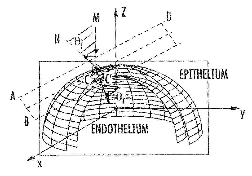
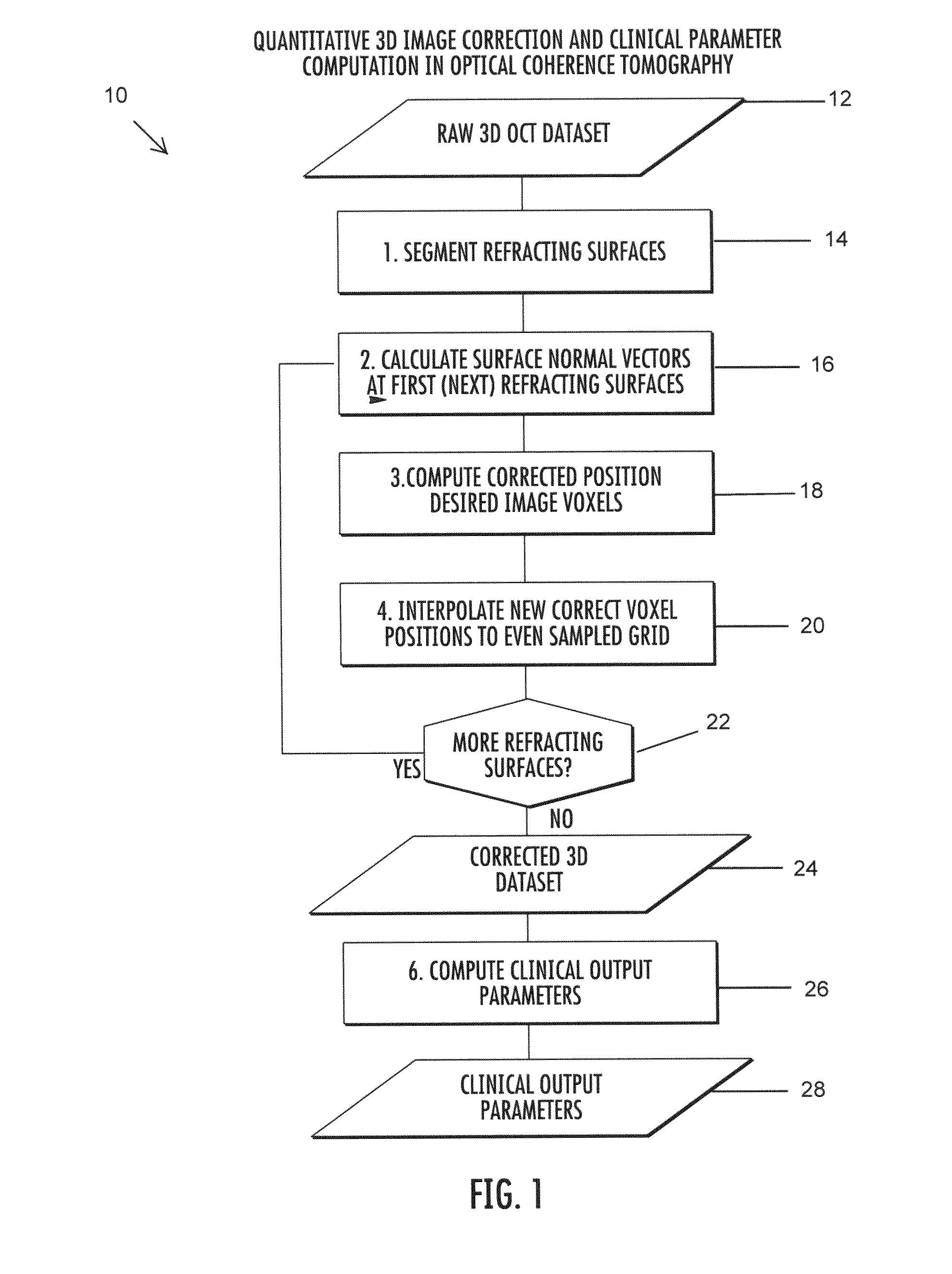
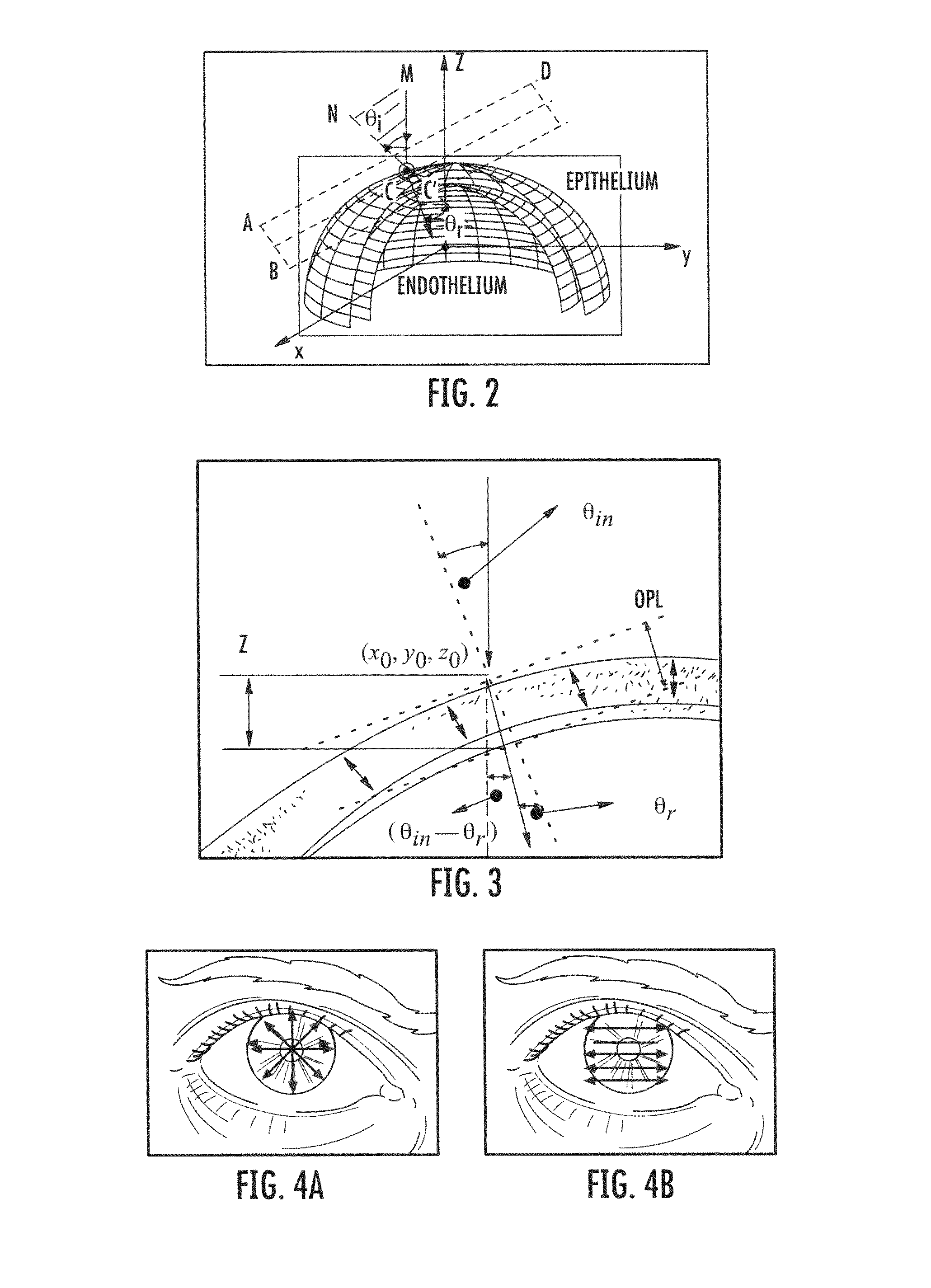
For meaningful analysis of the cornea of an eye from spectral domain optical coherence tomography (“SD-OCT”) data, effects of refraction at the epithelial and endothelial surfaces should be accounted for. A method for three-dimensional (“3D”) refraction correction based on a vector representation which accounts for refraction of optical coherence tomography (“OCT”) light in the cornea is present herein below. This method can be used to dewarp raw SD-OCT volumes. The method can also be used to reconstruct the tru...


PUM


Abstract

Methods and computer program products for quantitative three-dimensional (“3D”) image correction in optical coherence tomography. Using the methods and computer program products, index interface (refracting) surfaces from the raw optical coherence tomography (“OCT”) dataset from an OCT system can be segmented. Normal vectors or partial derivatives of the curvature at a refracting surface can be calculated to obtain a refracted image voxel. A new position of each desired refracted image voxel can be iteratively computed. New refracted corrected voxel positions to an even sampling grid can be interpolated to provide corrected image data. In some embodiments, clinical outputs from the corrected image data can be computed.

Description

TECHNICAL FIELDThe present subject matter generally relates to optical coherence tomography (“OCT”). More particularly, the present subject matter relates to OCT for use in analysis of ocular optical elements and surfaces, such as the cornea and lens of the eye.BACKGROUNDMultiple modalities currently exist to image the ocular anterior segment of an eye. These imaging devices aid in the clinical diagnosis and care of ocular disease. Optical coherence tomography (“OCT”) is one such technique used to image in vivo the ocular anterior segment of an eye in a non-invasive fashion. Specific implementations can comprise time-domain OCT systems (such as those sold under the name of VISANTE® by Carl Zeiss Meditec, Inc.), and Fourier-domain OCT systems, including both swept-source and spectrometer-based spectral-domain (“SDOCT”) implementations. Many previously disclosed anterior segment OCT systems have utilized illumination light in the 1310 nm region. Recently, several SD-OCT systems have b...

Claims


Application Information

Patent Timeline
Patent Type & Authority Applications(United States)
IPC IPC(8): G01B11/02
CPCG01B11/2441G01B9/02091G01B9/02083G06T2207/30041G06T15/00G06T2207/10101G06T5/006A61B3/102
Inventor IZATT, JOSEPH A.ZHAO, MINGTAOKUO, ANTHONY N.
Owner DUKE UNIV
Why Eureka
Try Eureka