Methods for treating demyelination disorders
a demyelination disorder and demyelination technology, applied in the field of neurology, can solve the problems of inability to cure and few effective therapies, lack of understanding of the precise mechanism by which the secretion of ifn- leads to oligodendroglial abnormalities, and inability to achieve a sustained and reduce the stress level of endoplasmic reticulum. er
- Summary
- Abstract
- Description
- Claims
- Application Information
AI Technical Summary
Benefits of technology
Problems solved by technology
Method used
Image
Examples
example 1
IFN-γ Does not Affect the Initial Phase Demyelination, Oligodendrocyte Loss or Reduction of Myelin Gene Expression
[0165] The effects of IFN-γ on demyelination were evaluated in a cuprizone animal model using transgenic mice that allow for temporally regulated delivery of IFN-γ using the tetracycline controllable system (Lin, et al. (2004) J. Neurosci. 24: 10074-10083). The transgenic mice were generated by mating line 110 GFAP / tTA mice on the C57BL / 6 background with line 184 TRE / IFN-γ on the C57BL / 6 background to produce GFAP / tTA; TRE / IFN-γ double transgenic mice (Lin, et al. (2004) J. Neurosci. 24: 10074-10083). Transcriptional activation of the TRE / IFN-γ transgene by tTA was repressed in the control (DOX+) mice by adding 0.05 mg / ml doxycycline to the drinking water which was provided ad libitum from the day of conception. The DOX+ double transgenic mice were GFAP / tTA; TRE / IFN-γ double transgenic animals fed cuprizone chow and never released from the doxycycline solution. DOX− dou...
example 2
IFN-γ Suppresses Remyelination in Demyelinated Lesions
[0178] The effect of IFN-γ on the remyelination in the cuprizone treated mice described in Example 1 was determined following withdrawal of cuprizone at week 5. The experimental methods used are the same as described in Example 1.
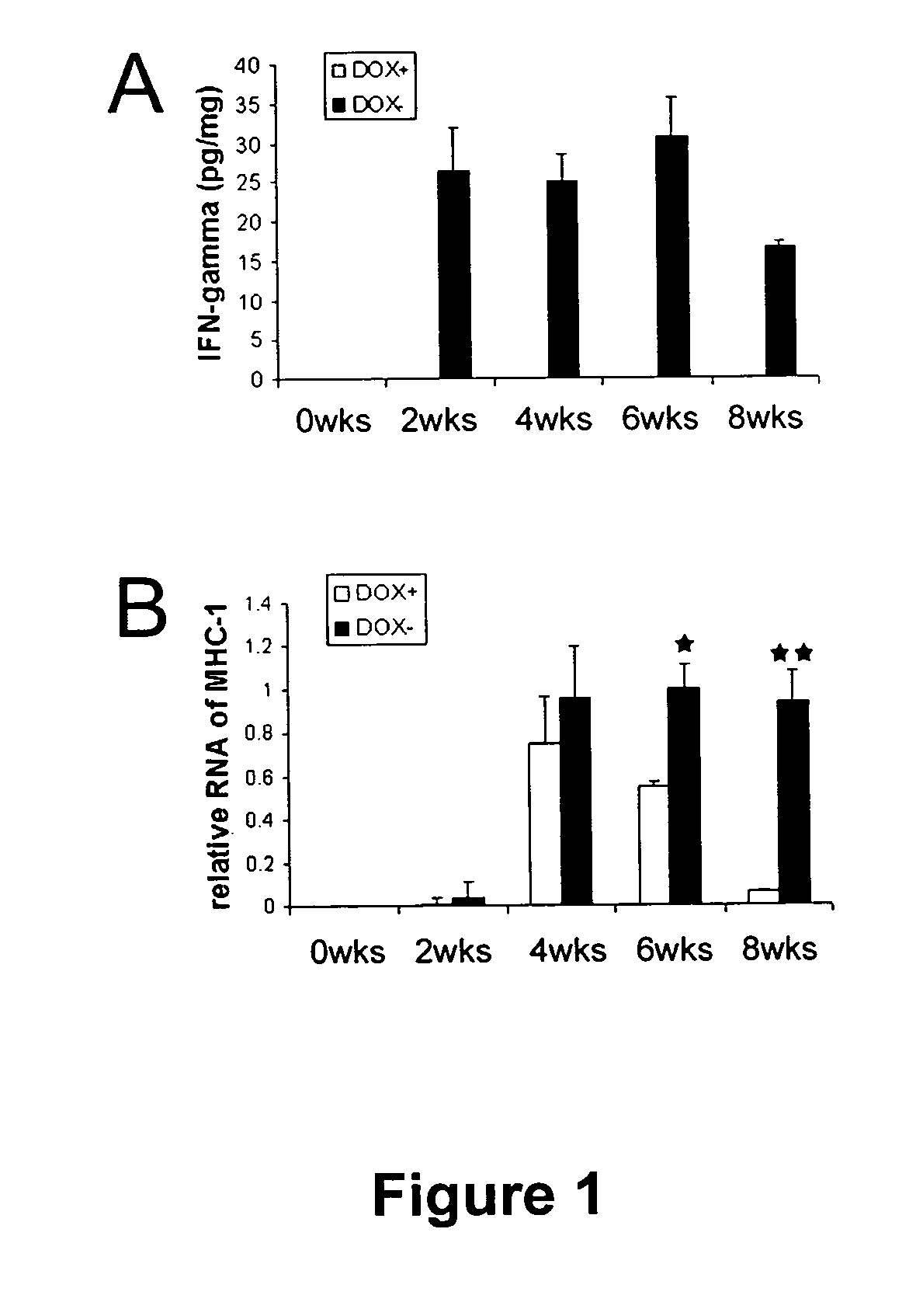
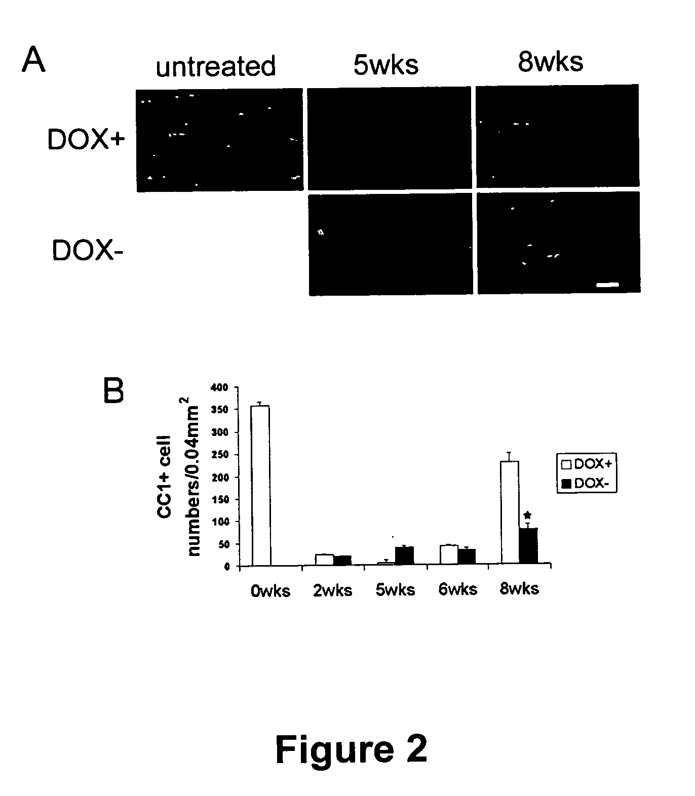
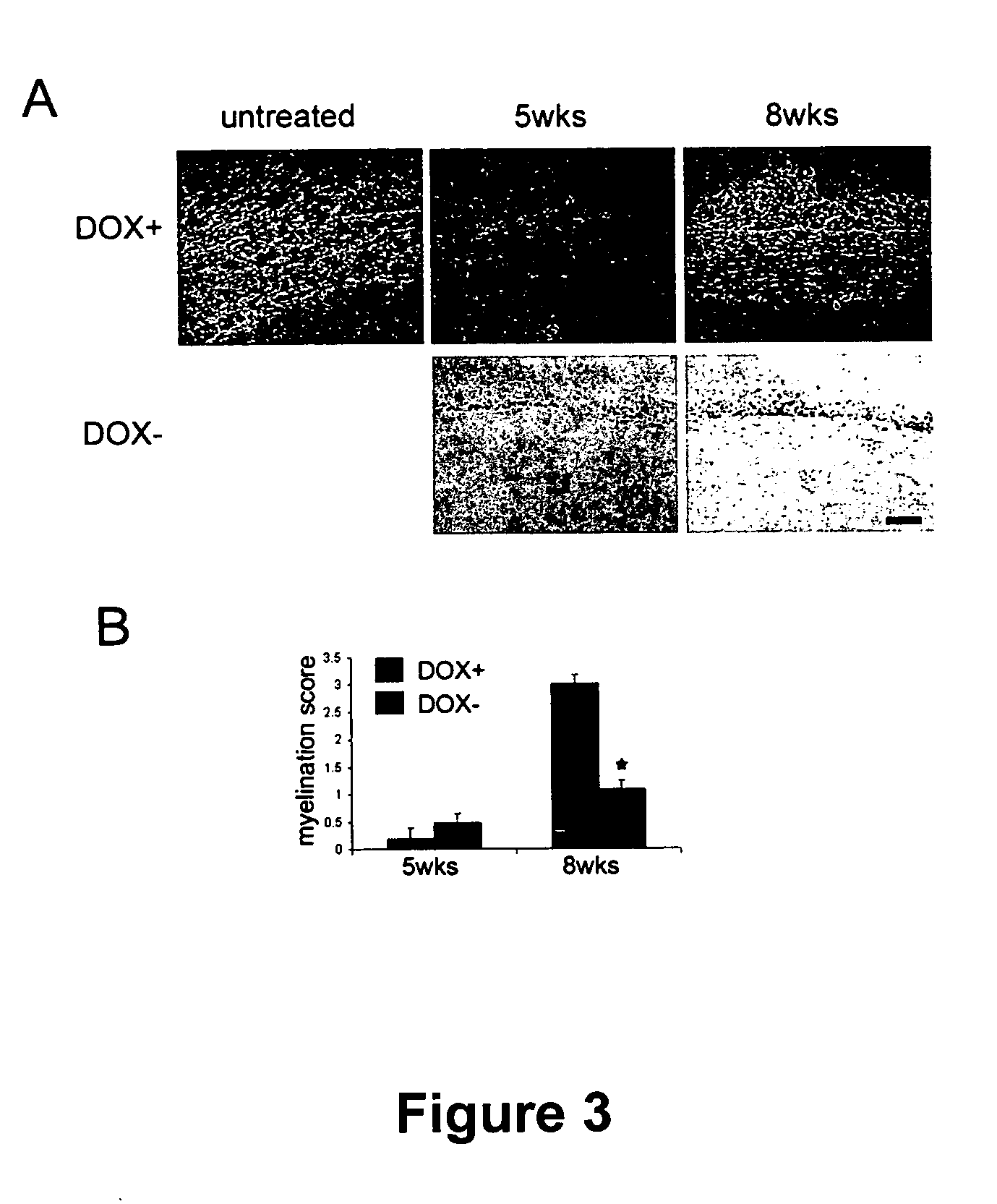
[0179]FIGS. 1A and B show that the increased level of INF-γ persisted in the DOX− mice even after withdrawal of cuprizone, and the increase in INF-γ was accompanied by a sustained increase in MCH-1. By week 8, a significant number of oligodendrocytes were seen in the corpus callosum of the DOX+ mice, while the regeneration of oligodendrocytes in the DOX− was suppressed (FIGS. 2A and B). Remyelination in the corpus callosum of DOX+ double transgenic mice began at week 6, and was evident 2 weeks after cuprizone was removed from the diet (week 8; FIG. 3). In control mice DOX+, a large number of axons showed substantial recovery at 8 weeks (48.4%±4%, FIG. 4), whereas remyelination remained markedly suppres...
example 3
IFN-γ Dramatically Reduces Repopulation of CC1 Positive Oligodendrocytes in Demyelinated Lesions
[0181] Remyelination occurs by the repopulation of demyelinated lesions by oligodendrocyte precursors (OPCs) the site of the lesion where they differentiate into oligodendrocytes (Matsushima, et al. (2001) Brain Pathol. 11: 107-116; Mason, et al. (2000) J. Neurosci. Res. 61: 251-262). CC1 positive oligodendrocytes are derived from NG2 positive OPCs (Watanabe et al 1998; Mason et al 2000)). Therefore, repopulation of a lesion may be evaluated by the presence of CC1 and / or NG2 positive cells.
[0182] The effect of INF-γ on the repopulation of demyelinated lesions by oligodendrocyte precursors (OPC) was evaluated in the mice of Example 2 as the number of CC1 positive mature oligodendrocytes and NG2 positive OPCs that were counted in sections from the corpus callosum of both DOX+ and DOX− mice. The methods used for analyzing the oligodendrocytes during remyelination is the same as that descri...
PUM
| Property | Measurement | Unit |
|---|---|---|
| Fraction | aaaaa | aaaaa |
| Time | aaaaa | aaaaa |
| Time | aaaaa | aaaaa |
Abstract
Description
Claims
Application Information
 - R&D
- Intellectual Property
- Life Sciences
- Materials
- Tech Scout
- Unparalleled Data Quality
- Higher Quality Content
- 60% Fewer Hallucinations
Browse by: Latest US Patents, China's latest patents, Technical Efficacy Thesaurus, Application Domain, Technology Topic, Popular Technical Reports.
© 2025 PatSnap. All rights reserved.Legal|Privacy policy|Modern Slavery Act Transparency Statement|Sitemap|About US| Contact US: help@patsnap.com


