
X-ray computed tomography apparatus

a computed tomography and x-ray technology, applied in the direction of material analysis using wave/particle radiation, instruments, applications, etc., can solve the problems of time resolution, image quality inevitably deteriorating, and it is difficult to design an optimal cardiac phas

Inactive Publication Date: 2007-02-08
TOSHIBA MEDICAL SYST CORP
View PDF16 Cites 27 Cited by
  • Summary
  • Abstract
  • Description
  • Claims
  • Application Information

AI Technical Summary

Benefits of technology

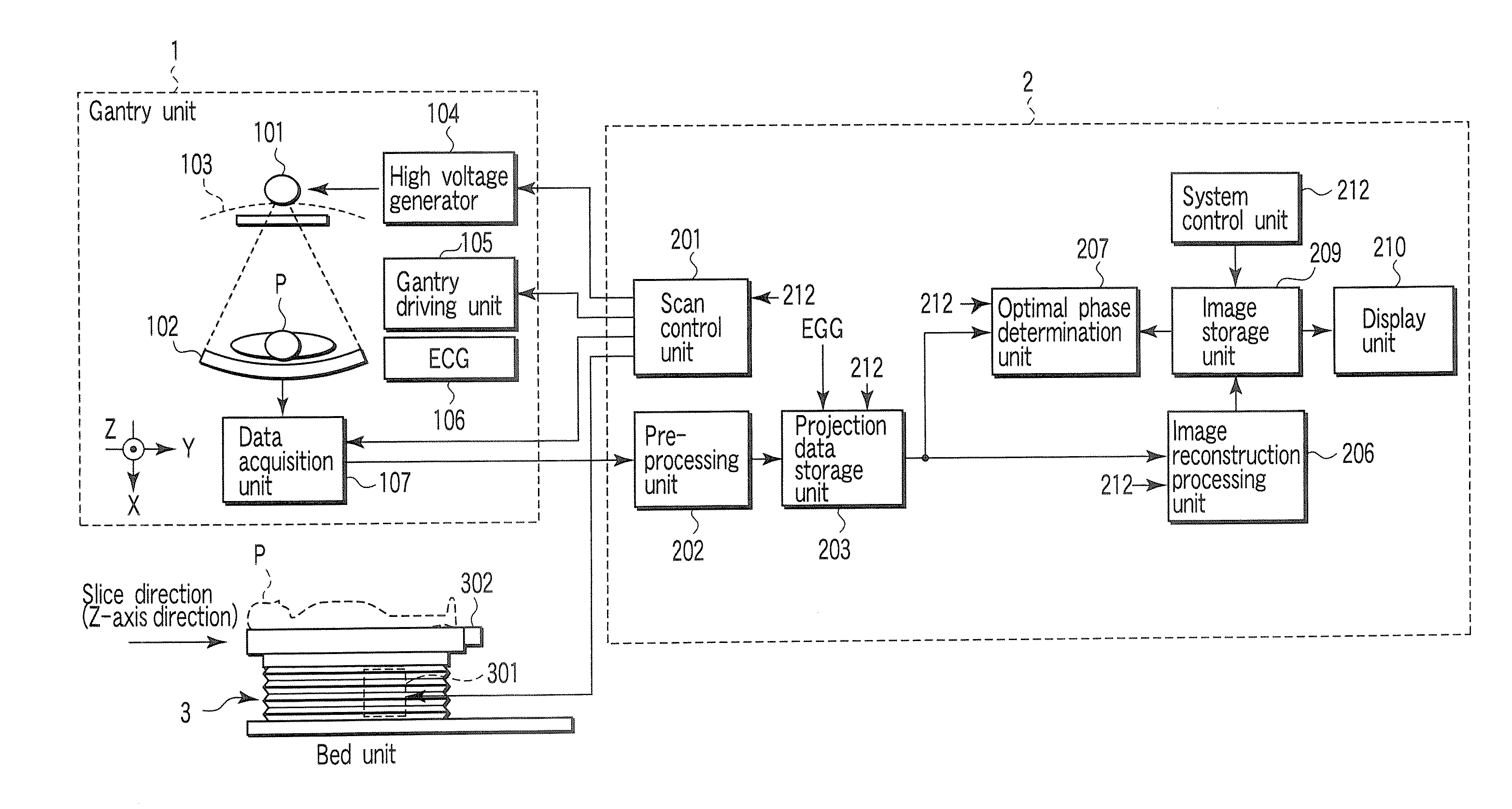
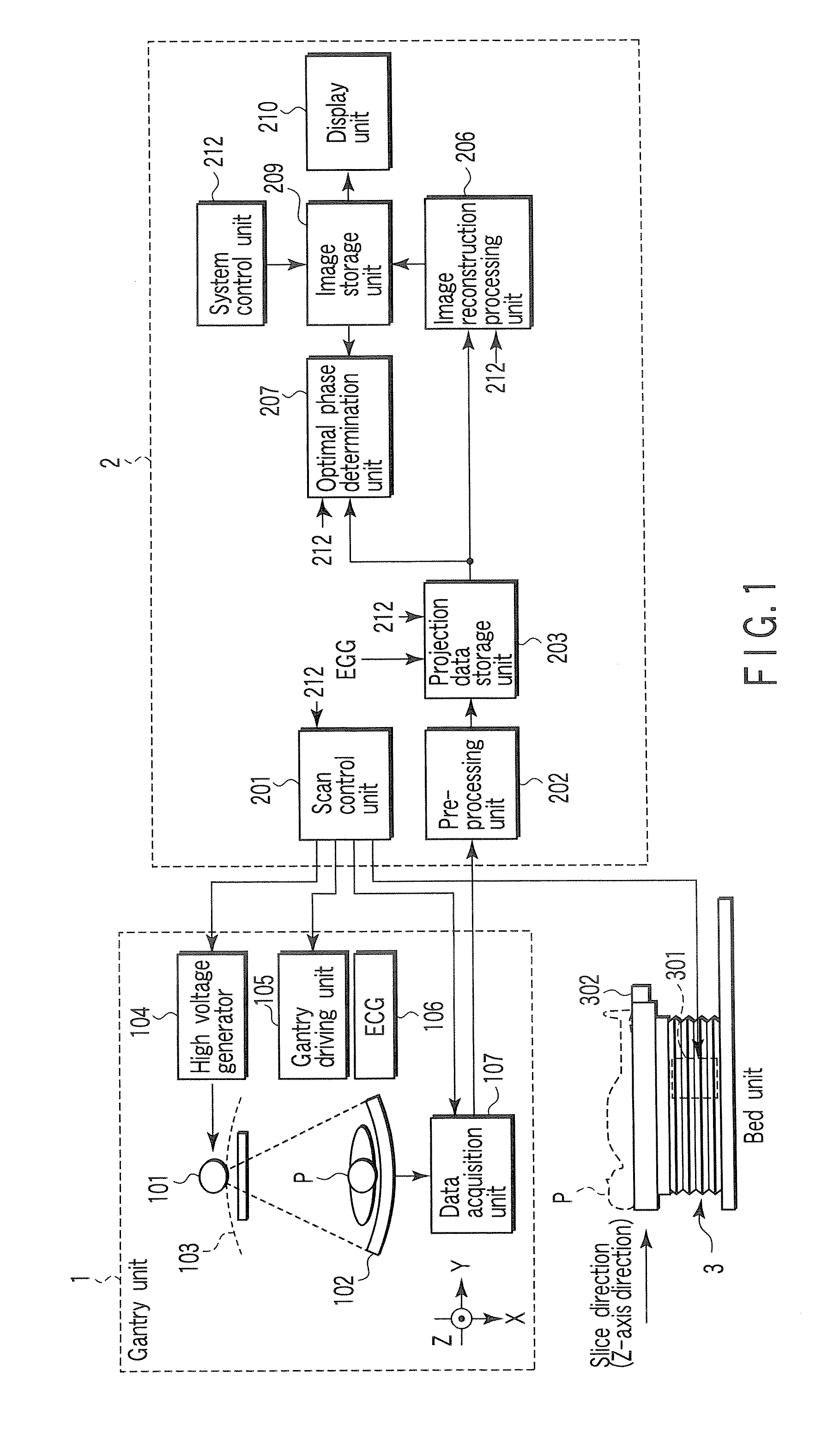
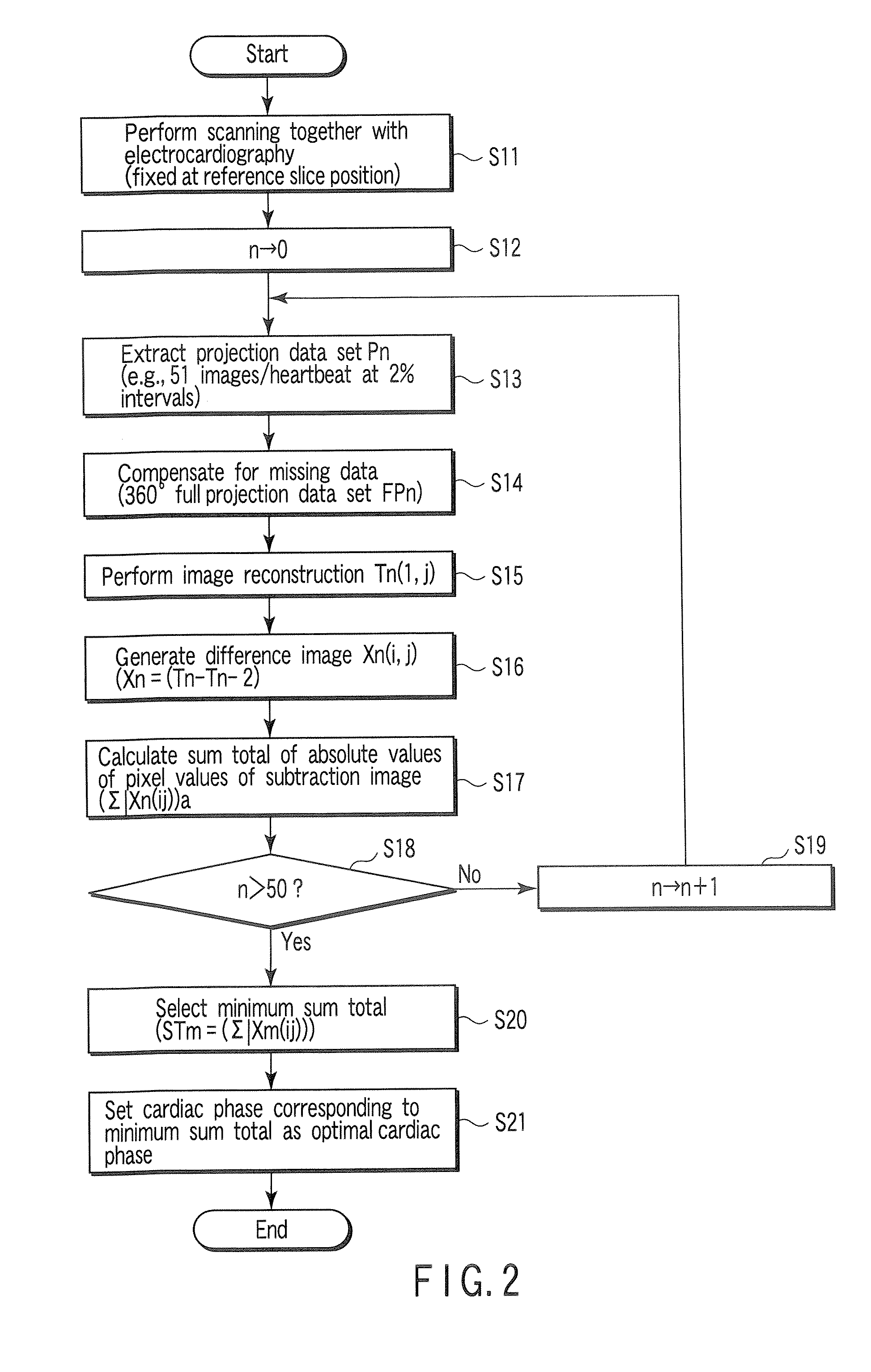
[0012] The accompanying drawings, which are incorporated in and constitute a part of the specification, illustrate embodiments of the invention, and together with the general description given above and the detailed description of the embodiments given below, serve to explain the principles of the invention.

Problems solved by technology

In examination of a fast-moving part using an X-ray computed tomography apparatus, and in particular, cardiac examination, one of the important challenges is to improve the time resolution of images.
Therefore, image quality inevitably deteriorates due to blurring or the like originating from the magnitude of the pulsation of the heart within this substantial time resolution.
In many cases, it has been difficult to designate an optimal cardiac phase, i.e., a cardiac phase exhibiting the least movement of the heart in a time width which corresponds to the substantial time resolution and is centered on the cardiac phase (see Jpn. Pat. Appln. KOKAI Publication No. 2004-275440).

Method used


Image

  • X-ray computed tomography apparatus
  • X-ray computed tomography apparatus
  • X-ray computed tomography apparatus

Examples


Embodiment Construction

[0028] An embodiment of an X-ray computed tomography apparatus according to the present invention will be described below with reference to the views of the accompanying drawing. Note that X-ray computed tomography apparatuses include various types of apparatuses, e.g., a rotate / rotate-type apparatus in which an X-ray tube and X-ray detector rotate together around a subject to be examined, and a stationary / rotate-type apparatus in which many detection elements are arrayed in the form of a ring, and only an X-ray tube rotates around a subject to be examined. The present invention can be applied to either type. In this case, the rotate / rotate type, which is currently the mainstream, will be exemplified. In order to reconstruct one-slice tomogram data, a full projection data set (full reconstruction method) corresponding to one rotation around a subject to be examined, i.e., about 360°, is required, or a half projection data set corresponding to 180°+α (α: fan angle) is required in the...


PUM


Abstract

An X-ray computed tomography apparatus includes an X-ray tube which generates X-rays, an X-ray detector which detects X-rays transmitted through a subject to be examined, a mechanism which continuously rotates the X-ray tube and the X-ray detector, a storage unit which stores projection data detected by the X-ray detector, a read unit which reads out projection data sets of parts of a pair spaced apart from each other by 360° from the storage unit, an index generating unit which generates a plurality of indices indicating movement of a heart on the basis of a difference between the projection data sets of the parts of the pair, a cardiac phase determination unit which determines a cardiac phase on the basis of the indices, and a reconstruction unit which reconstructs an image on the basis of a full projection data set corresponding to the determined cardiac phase.

Description

CROSS-REFERENCE TO RELATED APPLICATIONS [0001] This application is based upon and claims the benefit of priority from prior Japanese Patent Application No. 2005-225537, filed Aug. 3, 2005, the entire contents of which are incorporated herein by reference. BACKGROUND OF THE INVENTION [0002] 1. Field of the Invention [0003] The present invention relates to an X-ray computed tomography apparatus which scans a subject to be examined with X-rays and reconstructs image data on the basis of obtained projection data using an ECG-gated reconstruction method. [0004] 2. Description of the Related Art [0005] An X-ray computed tomography apparatus provides information about a subject to be examined in the form of images on the basis of the intensities of X-rays transmitted through the subject, and plays important roles in many medical activities such as disease diagnosis and medical and operation planning. [0006] In examination of a fast-moving part using an X-ray computed tomography apparatus, ...

Claims


Application Information

Patent Timeline
IPC IPC(8): A61B6/00G01N23/00G21K1/12H05G1/60
CPCA61B6/541A61B6/503
Inventor TSUYUKI, MASAHARUOOTA, TAKAMASA
Owner TOSHIBA MEDICAL SYST CORP
Learn More