

Self-Assembling Cell Aggregates and Methods of Making Engineered Tissue Using the Same
a cell aggregate and self-assembling technology, applied in the field of tissue engineering, can solve the problems of lack of suitable organ replacement, many will likely perish, and the variety of tissue engineering methods and devices have been attempted and developed with limited success, and the assembly of vascularized three-dimensional soft organs has not been accomplished
- Summary
- Abstract
- Description
- Claims
- Application Information
AI Technical Summary
Benefits of technology
Problems solved by technology
Method used
Image

Examples
example 1
Cell Aggregate Preparation
[0099]Chinese Hamster Ovary (CHO) cells, transfected with N-cadherin (courtesy of A. Bershadsky, Weizmann Institute, Rehovot, Israel), were infected with histone binding H2B-YFP retrovirus (provided by R. D. Lansford, Beckman Institute at California Institute of Technology). Confluent cell cultures (3-4×10⁶ cells / 75 cm2 TC dish) grown in Dulbecco's Modified Eagle Medium (DMEM, Gibco BRL Grand Island, N.Y.; supplemented with 10% FES (US Biotechnologies, Parkerford, Pa.), 10 μg / ml of penicillin, streptomycin, gentamicin, kanamycin, 400 μg / ml geneticin), were washed twice with Hanks' Balanced Salt Solution (HBSS) containing 2 mM CaCl2, then treated for 10 minutes with trypsin 0.1% (diluted from 2.5% stock, Gibco BRL, Grand Island, N.Y.). Depleted cells were centrifuged at 2500 RPM for 4 minutes. The resulting pellet was transferred into capillary micropipettes of 500 μm diameter and incubated at 37° C. with 5% CO2 for 10 minutes. The firm cylinders of cells re...
example 2
Formation of a Fused Ring Structure
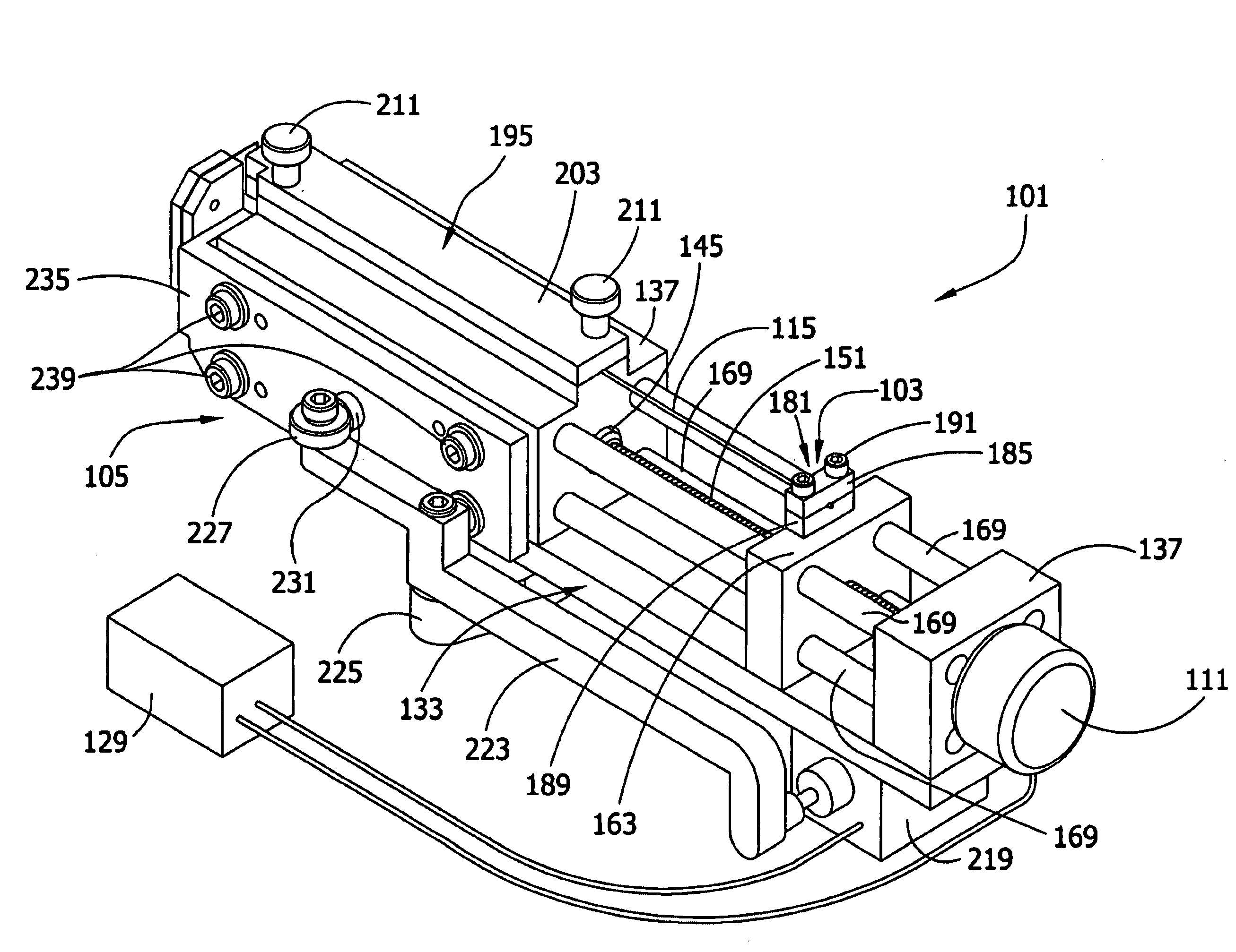
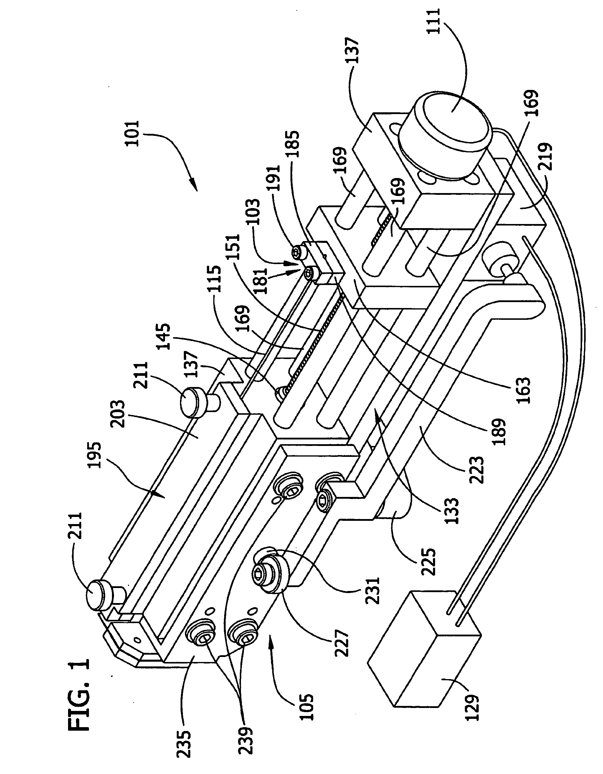
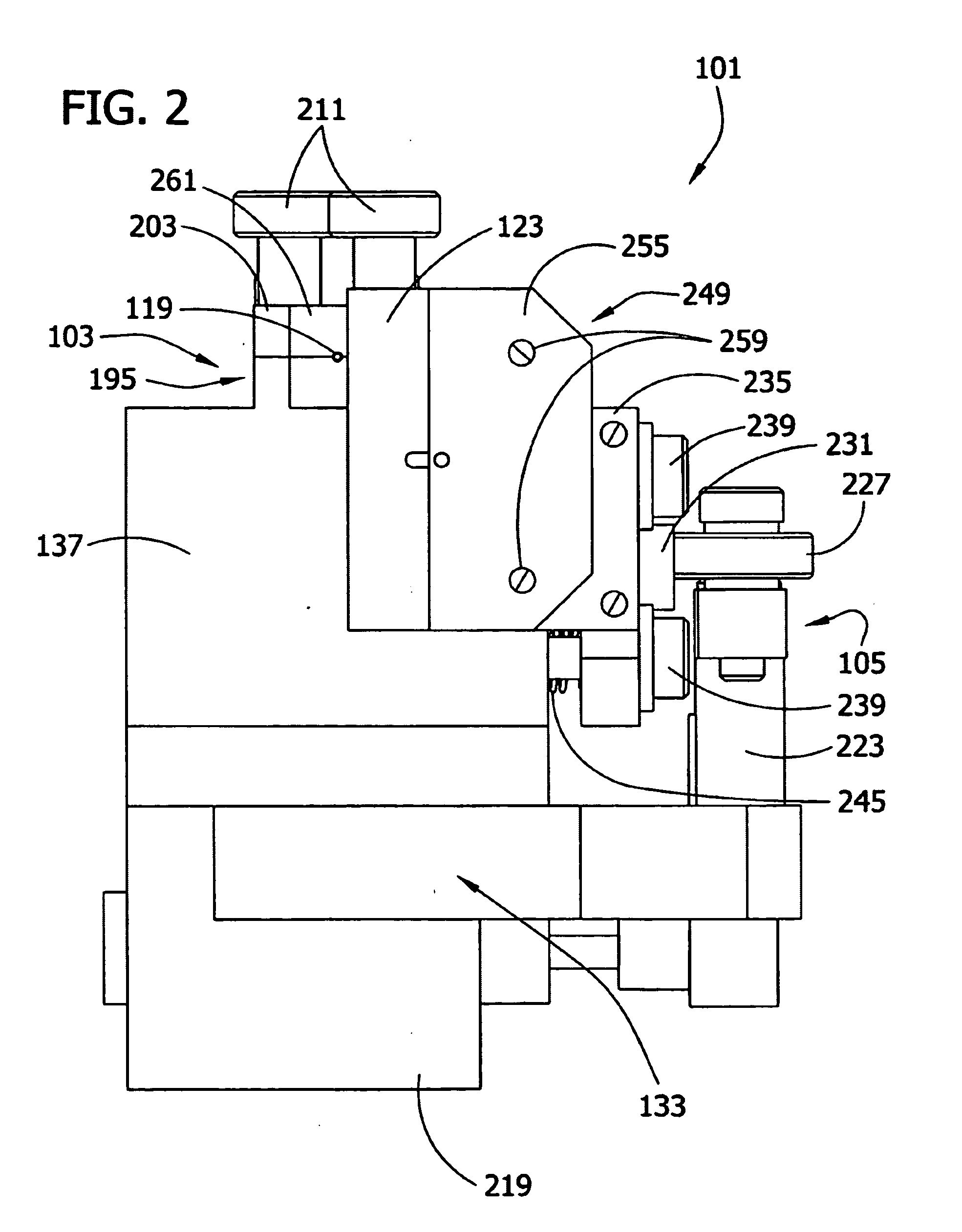
[0100]The cell aggregates prepared in Example 1 were used to engineer a three dimensional tissue construct by either “manually printing” or using computer controlled delivery devices to print (i.e., embedding) the aggregates into biocompatible gels in a predetermined pattern.
[0101]NeuroGel™ (a biocompatible porous poly[N-(hydroxypropyl)methacrylamide] hydrogel) disks of 10 mm diameter and 2 mm thickness, containing RGD fragments (provided by Stephane Woerly, Organogel Canada, Quebec) were washed three times with DMEM to eliminate the storage medium. A 0.5 mm wide, 0.5 mm deep circular groove was cut into a disk. Ten aggregates were placed (either manually or by printing using a device as described herein) contiguously in the groove to form a closed circle. The groove was refilled with the gel to completely embed the aggregates. This structure was incubated at 37° C., 5% CO2 for 72 hours in a tissue culture dish containing 10 ml DMEM, washed with PB...
example 3
Identification of a Long-Lived Structure
[0108]Shapes corresponding to long-lived structures (e.g., toroidal structures), may be identified from plateaus in the plot of the total interaction energy vs. MCS. This is illustrated in more detail in the simulation shown in FIG. 11A and FIG. 11B (γcg / ET=1.1), where the initial state progresses towards a long-lived toroidal configuration, whose energy is essentially unchanged in the entire interval between 104 and 6×10⁴ Monte Carlo steps (MCS) (FIG. 11A). Eventually the toroid becomes unstable, and at about 105 MCS it ruptures (FIG. 11B). Subsequent massive rearrangements lead to a pronounced energy decrease while the system evolves into three rounded aggregates. These remain stable for a long time because large spatial separations hinder their fusion into a single spheroid.
[0109]Once a structure reaches the long-lived state, it can be stabilized by dissolving the supporting gel or polymer. In the simulations this corresponds to increasing ...
PUM
| Property | Measurement | Unit |
|---|---|---|
| Fraction | aaaaa | aaaaa |
| Diameter | aaaaa | aaaaa |
| Length | aaaaa | aaaaa |
Abstract
Description
Claims
Application Information