Dental Optical Diagnostic Apparatus

a diagnostic apparatus and optical technology, applied in the field of diagnostic equipment, can solve the problems of difficult to clearly see dental carious, inability to confirm recalcification, and inability to define the threshold value uniformly

Inactive Publication Date: 2008-04-17
J MORITA TOKYO MFG CORP
View PDF12 Cites 35 Cited by
  • Summary
  • Abstract
  • Description
  • Claims
  • Application Information

AI Technical Summary

Problems solved by technology

However, the above listed items of diagnosis are accompanied by respective problems as described below.
[1] A dental carious (decayed tooth) on the surface of either of two adjacent teeth as detected and diagnosed by means of an X-ray image or visually, irradiating the site with light by means of a lamp accompanies a problem that the displayed image is not clear and hence it is difficult to clearly see the dental carious.
[2] A decalcification of dental enamel (initial dental carious) and a calcification (feasibility of re-calcification) as diagnosed by means of visual observation, palpation using an explorer or fluorometry using a laser-excited fluorometer involve a problem that a re-calcification cannot be confirmed in detail visually and by palpation and, while the result of the fluorometric measurement is displayed by a numeral between 1 and 100, the threshold value is not defined uniformly and hence is not highly reliable.
[3] When an initial carious of dentin and the progress thereof are diagnosed visually, by means of palpation using an explorer or fluorometry using a laser-excited fluorometer, it is not possible to entirely clarify the symptom and, while the result of the fluorometric measurement is displayed by a numeral between 1 and 100, the threshold value is not defined uniformly and hence is not highly reliable.
[5] When the profile of a periodontal pocket is diagnosed visually or by means of a multi-angular X-ray image, it is not possible to entirely clarify the symptom visually and a multi-angular X-ray image involves an invasion upon the patient.
[7] When the condition of an adhering calculus is diagnosed visually, it is not possible to entirely clarify the invisible areas such as the inside of a periodontal pocket and the diagnosis lacks reliability.
[9] For an operation of determining if a dental pulp is alive or dead and identifying and diagnosing the inflammatory part of a dental pulp for progress by means of X-rays and a laser Doppler blood flowmeter, the use of three-dimensional X-ray CT involves an invasion upon the patient and the apparatus for three-dimensional X-ray CT is complex and costly.
Additionally, a laser Doppler blood flowmeter is costly to install.
[10] When the number and the positions of dental roots are confirmed by means of three-dimensional X-ray CT, the operation involves an invasion upon the patient and the overall arrangement is complex and costly.
[11] When a crack and / or a partial loss of a dental root are diagnosed by means of three-dimensional X-ray CT, the operation also involves an invasion upon the patient and the overall arrangement is complex and costly.
However, in the field of dental treatment, the object of observation normally includes hard tissues such as dentin and enamel, soft tissues of the gingiva and periodontal tissues and the space available for the observation is limited in the oral cavity where the rows of teeth exist and the overall profile varies significantly from person to person.

Method used


Image

  • Dental Optical Diagnostic Apparatus
  • Dental Optical Diagnostic Apparatus
  • Dental Optical Diagnostic Apparatus

Examples


Embodiment Construction

[0056] It is possible to provide a non-invasive high resolution dental diagnostic apparatus for dental care that is different from X-ray apparatus that are conventional main diagnostic means by using an OCT apparatus as described above.

[0057] Now, the present invention will be described in greater detail by referring to the accompanying drawings that illustrate preferred embodiments of the invention.

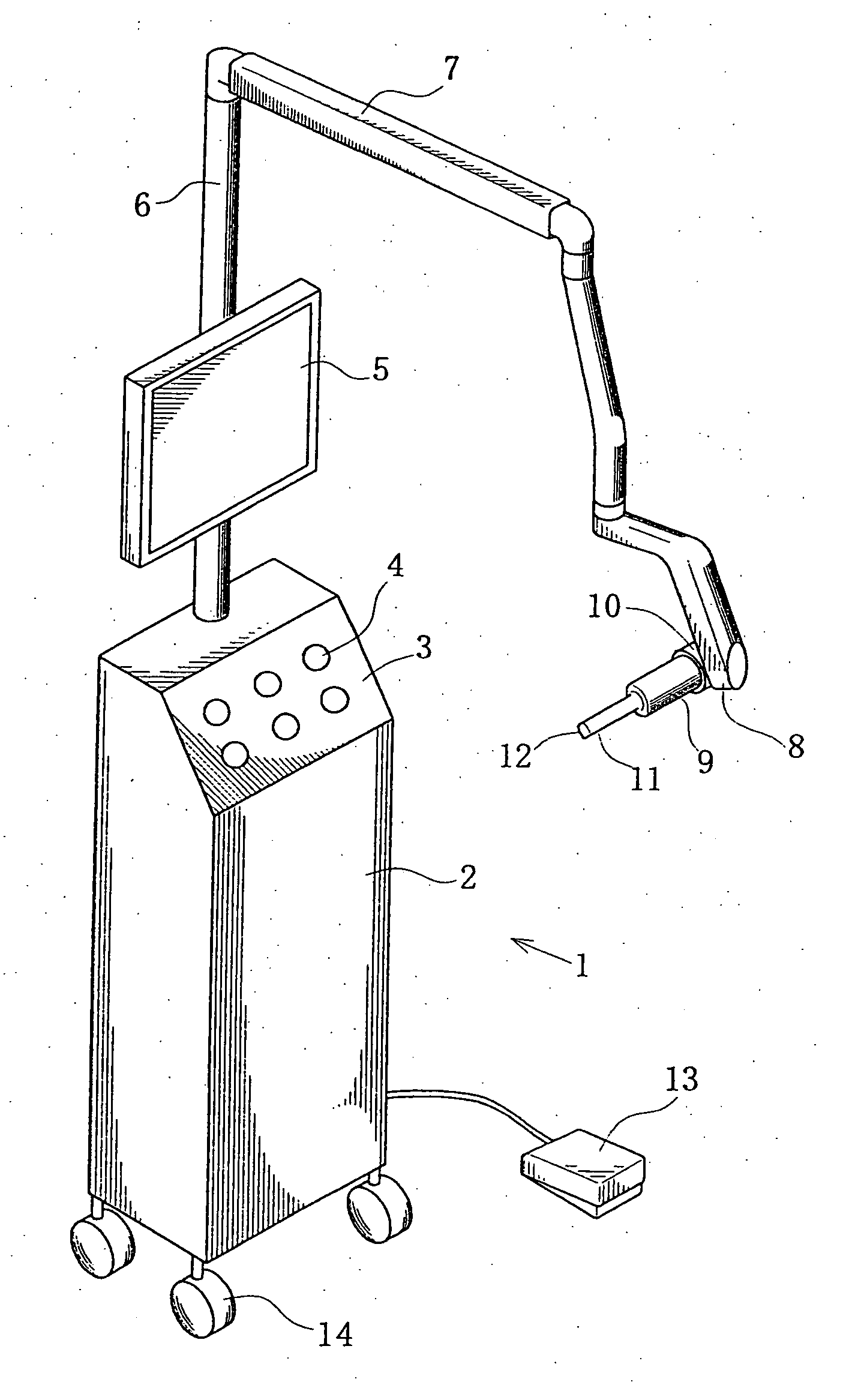
[0058]FIG. 1 is a schematic perspective view of a dental optical diagnostic apparatus according to the present invention and comprising an apparatus main body mounted on a cart and a diagnostic probe arranged at the front end of a multi-joint arm, showing the appearance thereof.

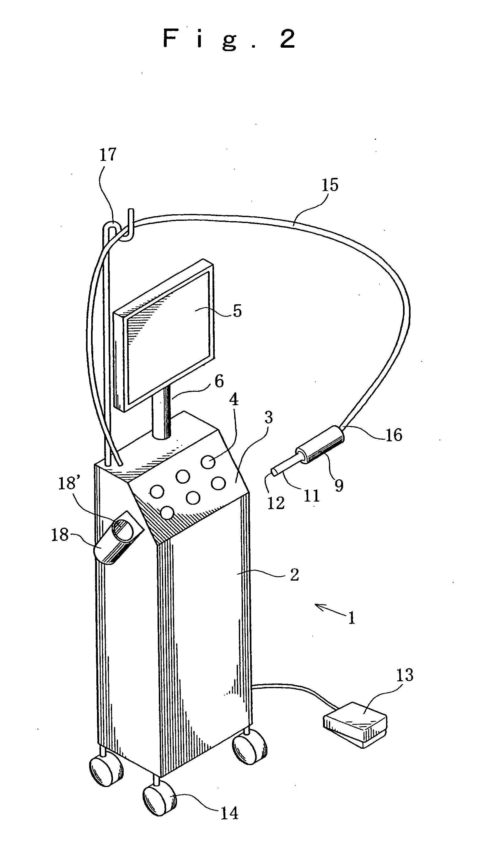
[0059]FIG. 1 shows an optical diagnostic apparatus 1 having a main body 2, an operation section 3, an operation switch 4, a display section 5, a pole 6, a multi-joint arm 7, a pivot 8 at the front end of the arm 7, a diagnostic probe 9, a pivot 10 of the probe 9, the front end 11 of the probe 9, an observation ...


PUM


Abstract

In the field of dental care, diagnoses have been and being made by means of X-ray images, visual observation, explorer, palpation, fluorometery, insertion of a pocket probe and observation, observation by means of a root canal meter and the use of a Doppler flood flowmeter. However, such diagnoses are invasive and not very clear and three-dimensional X-ray CT apparatus that are being used for diagnoses are very large and complex. Additionally, OCT apparatus for obtaining optical tomographys by using reflection light that are being used by ophthalmologists give rise to problems because of the profile of the detection probe and operability when such an apparatus is used in an oral cavity that contains hard tissues such as teeth and soft tissues such as gingiva for dental care. The present invention dissolves the above identified problems and provides a non-invasive and high resolution dental optical diagnostic apparatus. A dental optical diagnostic apparatus (1) according to the invention comprises a means (30) for generating guide light to irradiate the dental part of a subject for viewing, visible light for picking up a surface image and signal light of low coherence light for diagnosing the dental part, a camera (69) for picking up a surface image of the dental part, an OCT means for acquiring an optical tomography and an image display section (5) for displaying the obtained images.

Description

TECHNICAL FIELD [0001] This invention relates to a diagnostic apparatus in the field of dental care. More particularly, the present invention relates to an optical diagnostic apparatus using OCT (optical coherence tomography). BACKGROUND ART [0002] Conventionally, diagnostic apparatus and diagnostic methods have been and being appropriately and selectively used for diagnoses in the field of dental care according to the location and the symptom of the affected part. For instance, [1] A dental carious (decayed tooth) on the surface of either of two adjacent teeth is detected and diagnosed by means of an X-ray image or visually, irradiating the site with light by means of a lamp, [2] A decalcification of dental enamel (initial dental carious) and a calcification (feasibility of re-calcification) are diagnosed by means of visual observation, palpation using an explorer or fluorometry using a laser-excited fluorometer, [3] An initial carious of dentin and the progress thereof are diag...

Claims


Application Information

Patent Timeline
Patent Type & Authority Applications(United States)
IPC IPC(8): A61C3/00
CPCA61B5/0088A61B5/0066
Inventor NOGUCHI, KUNITOSHINISHIYAMA, FUMIOOGAWA, KAZUNOBUMIHATA, YUKINORISHIMAMURA, MASASHIKIMURA, SHUSUKE
Owner J MORITA TOKYO MFG CORP
Why Eureka
Try Eureka