Method and system for measuring left ventricle volume

a technology of left ventricle and volume measurement, applied in the field of medical imaging of the heart, can solve the problems of cardiac surgeons not being able to agree on the volume measurement field

Active Publication Date: 2009-03-26
SIEMENS HEALTHCARE GMBH
View PDF5 Cites 32 Cited by
  • Summary
  • Abstract
  • Description
  • Claims
  • Application Information

AI Technical Summary

Benefits of technology

[0005]The present invention provides a method and system for measuring left ventricle (LV) volume both including and excluding papillary muscles in a 3D cardiac CT volume. In order to determine volume measurements of the LV both including

Problems solved by technology

Cardiologists have not achieved agreement rega

Method used


Image

  • Method and system for measuring left ventricle volume
  • Method and system for measuring left ventricle volume
  • Method and system for measuring left ventricle volume

Examples


Embodiment Construction

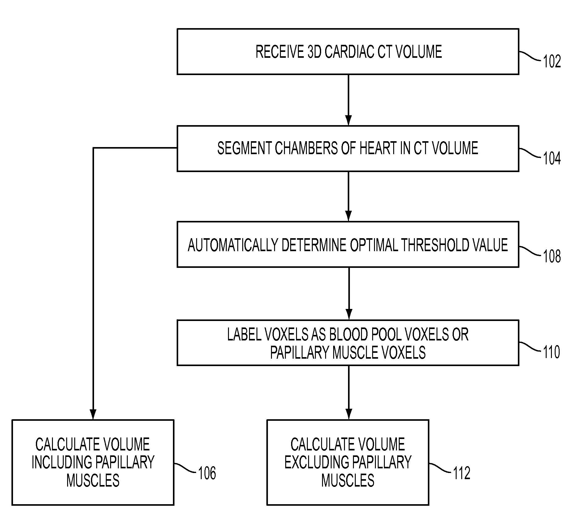
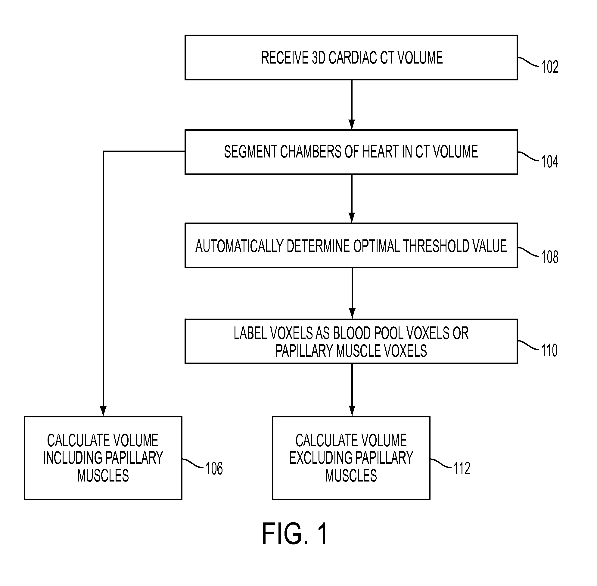
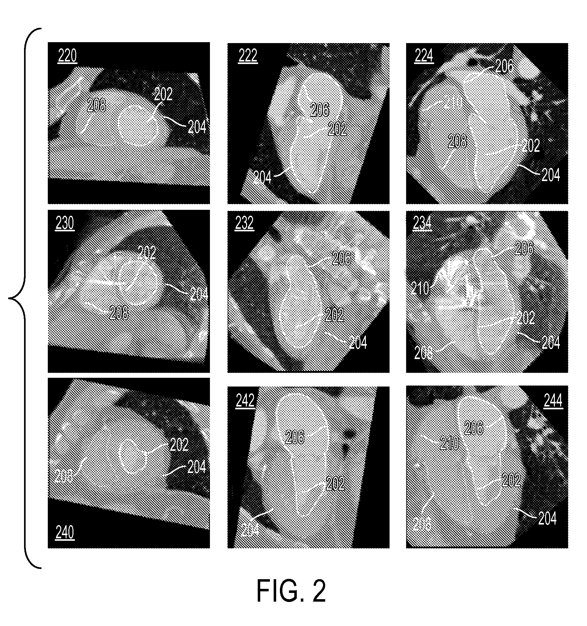
[0018]The present invention is directed to a method for measuring left ventricle (LV) volume in 3D medical images, such as computed tomography (CT) volumes, magnetic resonance images (MRI), and ultrasound images. Embodiments of the present invention are described herein to give a visual understanding of the heart modeling method. A digital image is often composed of digital representations of one or more objects (or shapes). The digital representation of an object is often described herein in terms of identifying and manipulating the objects. Such manipulations are virtual manipulations accomplished in the memory or other circuitry / hardware of a computer system. Accordingly, is to be understood that embodiments of the present invention may be performed within a computer system using data stored within the computer system.

[0019]Embodiments of the present invention measure LV volumes including and excluding the papillary muscles. Such embodiments of the present invention can be implem...


PUM


Abstract

A method and system for measuring the volume of the left ventricle (LV) in a 3D medical image, such as a CT, volume is disclosed. Heart chambers are segmented in the CT volume, including at least the LV endocardium and the LV epicardium. An optimal threshold value is automatically determined based on voxel intensities within the LV endocardium and voxel intensities between the LV endocardium and the LV epicardium. Voxels within the LV endocardium are labeled as blood pool voxels or papillary muscle voxels based on the optimal threshold value. The LV volume can be measured excluding the papillary muscles based on the number of blood pool voxels, and the LV volume can be measured including the papillary muscles based on the total number of voxels within the LV endocardium.

Description

[0001]This application claims the benefit of U.S. Provisional Application No. 60 / 974,139, filed Sep. 21, 2007, the disclosure of which is herein incorporated by reference.BACKGROUND OF THE INVENTION[0002]The present invention relates to medical imaging of the heart, and more particularly, to measuring a volume of the left ventricle of the heart in 3D cardiac CT volumes.[0003]The volume of a heart chamber and its dynamic change over the course of the heart cycle are important parameters in evaluating the functionality of the heart. The dynamic change of the volume of a heart chamber over the course of the heart cycle is typically measured as the ejection fraction (EF) of the heart chamber. Generally, the volume of a chamber at a given time can be determined by measuring the volume of the blood pool contained in the chamber. Typically, the volume of the left ventricle (LV) of the heart is determined in 3D CT volumes either by deformable model based automatic or semi-automatic methods ...

Claims


Application Information

Patent Timeline
IPC IPC(8): G06K9/00
CPCG06T7/0081G06T2207/30048G06T2207/10081G06T7/602G06T7/11G06T7/62
Inventor ZHENG, YEFENGGEORGESCU, BOGDANSCHEUERING, MICHAELCOMANICIU, DORIN
Owner SIEMENS HEALTHCARE GMBH
Why Eureka
Try Eureka