Catheter electrode that can simultaneously emit electrical energy and facilitate visualization by magnetic resonance imaging

a technology of electrical energy and electrodes, applied in the field of electrodes for catheters, can solve the problems of occupying valuable space and destroying tissue cells, and achieve the effect of retaining the ability to measure the flex of the catheter and reducing the number of tracking devices placed

Active Publication Date: 2009-07-02
ST JUDE MEDICAL ATRIAL FIBRILLATION DIV
View PDF83 Cites 36 Cited by
  • Summary
  • Abstract
  • Description
  • Claims
  • Application Information

AI Technical Summary

Benefits of technology

The patent text describes a new electrode design that can be used in MR imaging applications to directly measure the location of the electrode at the end of a catheter shaft. This design also maintains the electrical, thermal, and mechanical properties of conventional electrodes, while reducing the number of tracking devices placed on the shaft and allowing for the measurement of catheter flex.

Problems solved by technology

The technical problem addressed in this patent is the need for a way to directly measure the location of an electrode on a catheter, without relying on extrapolation or estimation, and to provide information about the flex of the catheter without having to place multiple tracking devices on the shaft.

Method used


Image

  • Catheter electrode that can simultaneously emit electrical energy and facilitate visualization by magnetic resonance imaging
  • Catheter electrode that can simultaneously emit electrical energy and facilitate visualization by magnetic resonance imaging
  • Catheter electrode that can simultaneously emit electrical energy and facilitate visualization by magnetic resonance imaging

Examples


Embodiment Construction

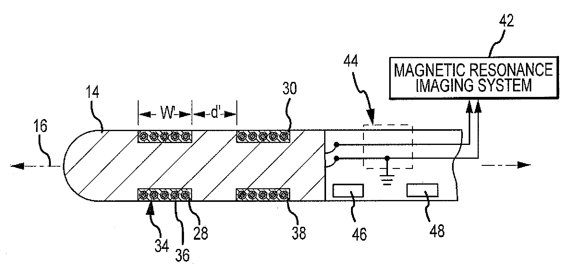
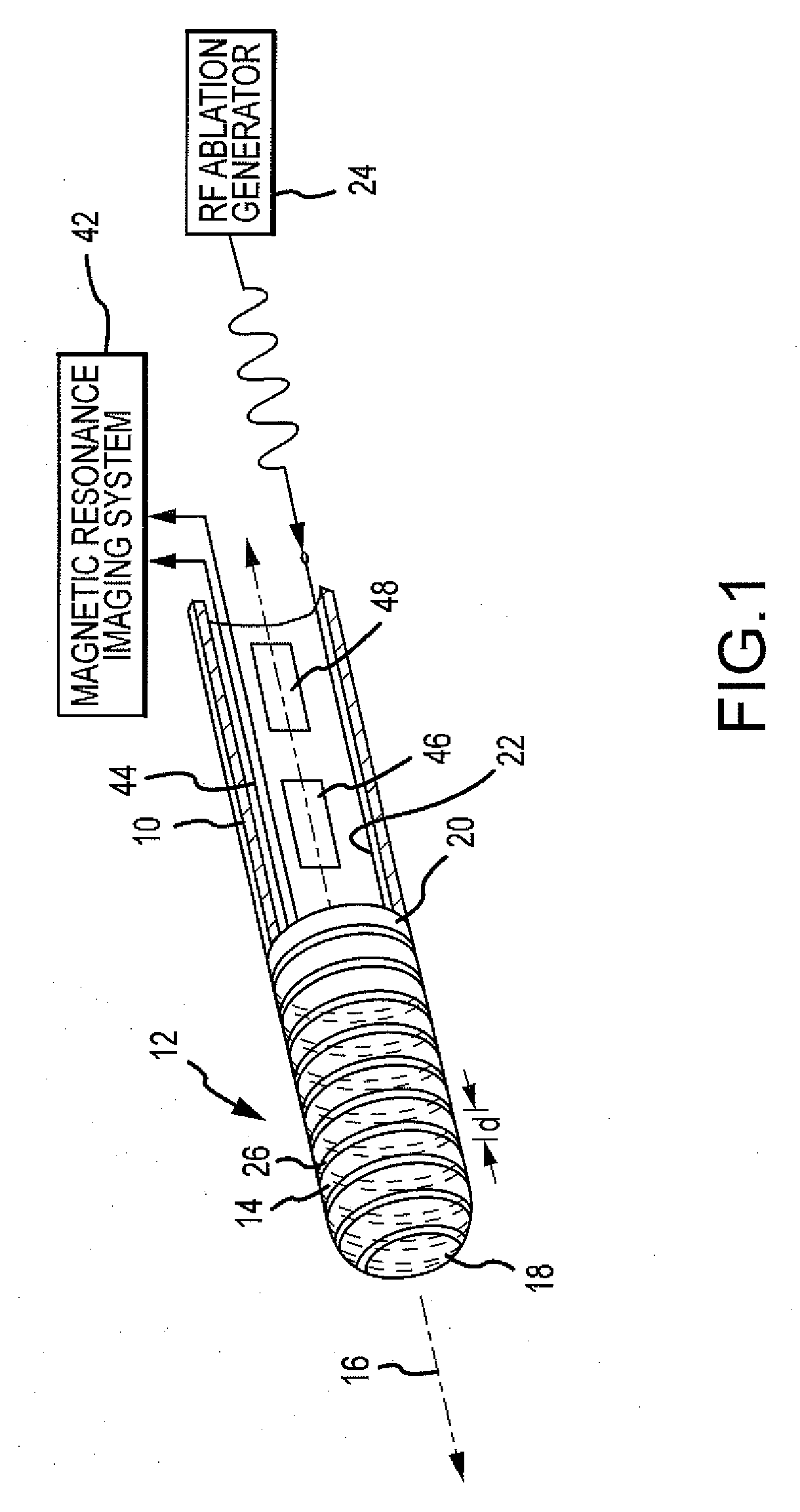
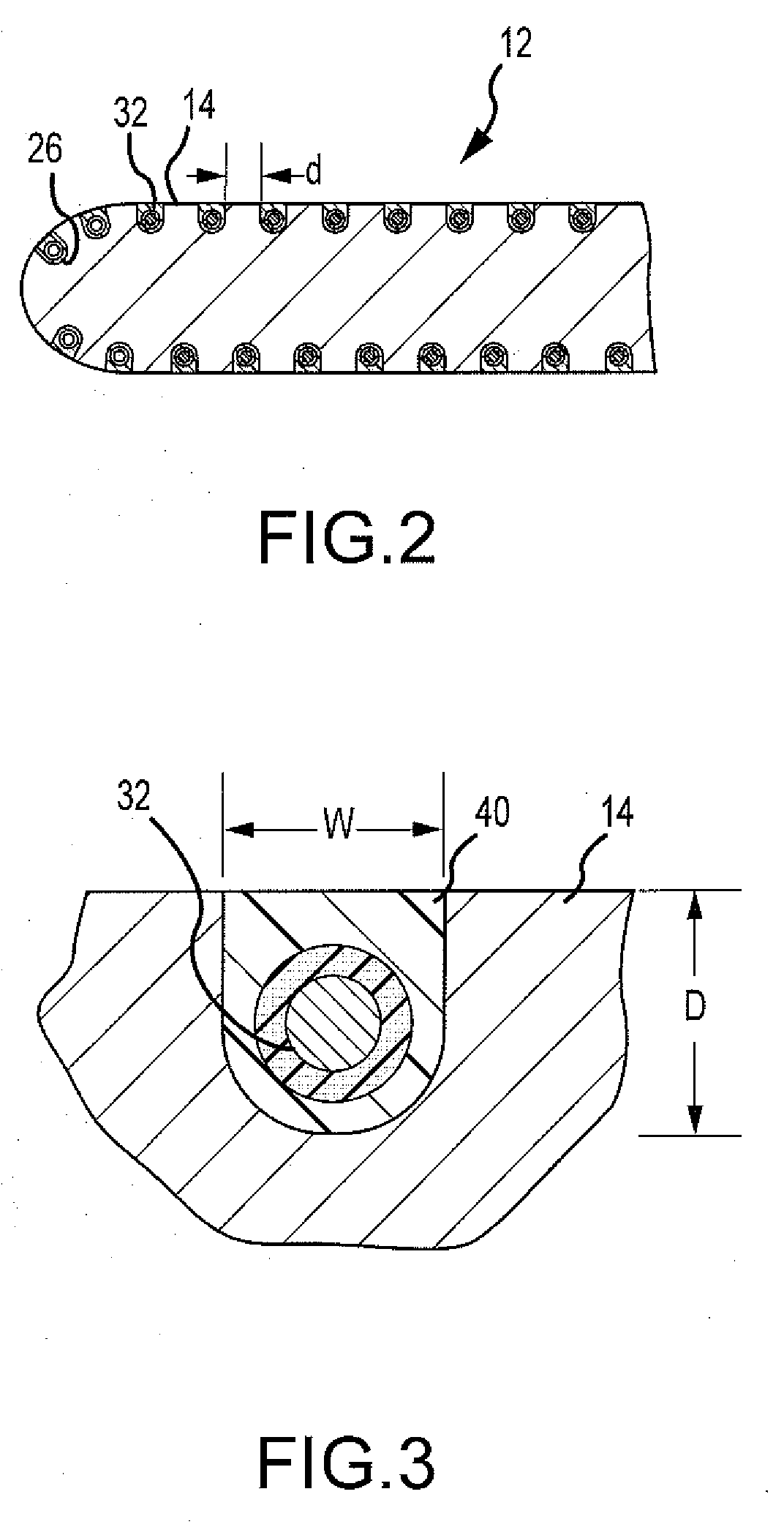
[0023]FIG. 1 illustrates a partial, simplified side view of a catheter including a shaft 10 with an electrode 12 in accordance with an embodiment of the invention. Shaft 10 may be designed for insertion into a main lumen of a sheath for eventual insertion into the body of a patient. Shaft 10 may comprise one or a plurality of layers. For example and without limitation, shaft 10 may comprise a braided layer of metal wire for additional stability and one or more layers of polymeric materials to create the desired stiffness and / or flexibility for the catheter. Shaft 10 may define one or more lumens for electrical leads, steering wires, or various other items that may be utilized within shaft 10. Shaft 10 may include a proximal section and a distal section. As used herein, “proximal” generally refers to a direction away from the body of a patient and toward a clinician. In contrast, “distal” generally refers to a direction toward the body of the patient and away from the clinician. Whil...


PUM


Abstract

An electrode for use on a medical device is disclosed. The electrode may have a main body of electrically conductive material extending along an axis and having a proximal end and a distal end. The body may be configured to emit electrical energy in accordance with a predefined diagnostic or therapeutic function. The body may have a groove disposed over an outermost surface of the body. The electrode may also include a magnetic resonance imaging (MRI) tracking coil disposed in said groove. The MRI tracking coil may comprise electrically insulated wire, for example. A catheter including an electrode, as well as a method for determining the location of an electrode, are also disclosed.

Description


Claims


Application Information

Patent Timeline
Owner ST JUDE MEDICAL ATRIAL FIBRILLATION DIV
Why Eureka
Try Eureka