
Enhanced transport using membrane disruptive agents

a technology of disruptive agents and transport channels, applied in drug compositions, microcapsules, metabolic disorders, etc., can solve problems such as disrupting the endosomal or other cellular membranes, and achieve the effect of enhancing endocytosis and preventing uptake and clearance of compositions

Inactive Publication Date: 2010-06-24
UNIV OF WASHINGTON
View PDF23 Cites 5 Cited by
  • Summary
  • Abstract
  • Description
  • Claims
  • Application Information

AI Technical Summary

Benefits of technology

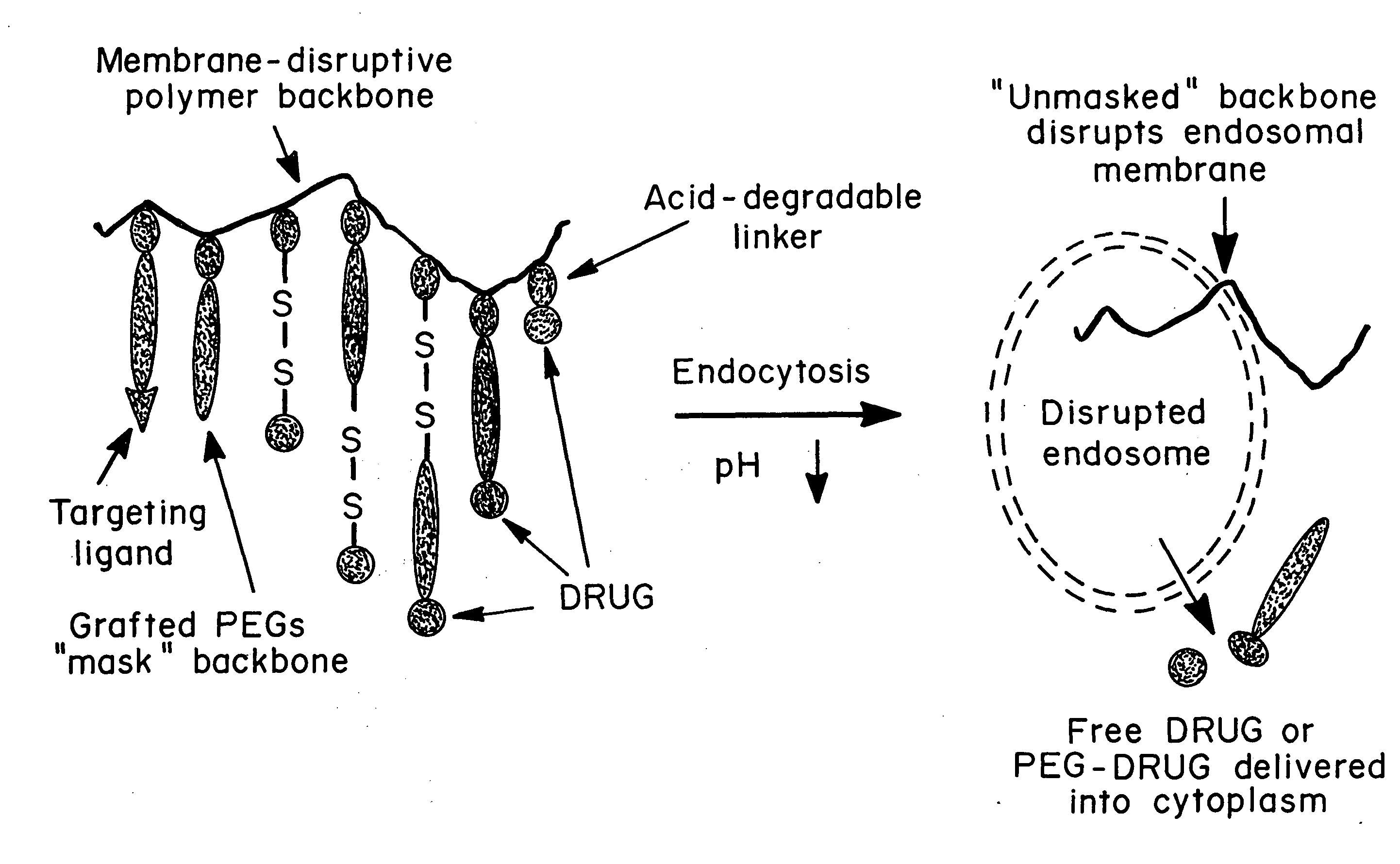
[0019]The hydrophilic groups can be linked directly to the hydrophobic polymeric backbone or can be on a polymer which is linked to the hydrophobic polymer via degradable (or disruptable) linkages. In either case, exposure to low pH or the stimulus which disrupts the linkages releases the hydrophobic polymeric backbone, which then disrupts the endosomal or other cellular membrane. In a preferred embodiment, the hydrophilic groups are a polymer, such as polyethylene glycol (PEG), which helps prevent uptake and clearance of the composition by phagocytic cells of the reticuloendothelial system (RES) prior to uptake by the cells to which the therapeutic, diagnostic or prophylactic agent is to be delivered. In another embodiment, the hydrophilic groups are PEG groups, which are conjugated directly to drug molecules and are conjugated to the hydrophobic backbone by acid degradable linkages (also referred to herein as “pH-degradable linkages”). In another embodiment, the hydrophilic polymer contains disulfide-linked drugs, designed to deliver drug-SH molecules within the cytoplasm.
[0020]The membrane disruptive agents may be used to deliver “fragile” drugs, such as DNA plasmids, antisense oligodeoxynucleotides (ODNs) and cytoplasmic-based protein therapeutics, as immunotoxins. They may serve as carriers for proteins or peptides designed for entry into the MHC 1 pathway for vaccine applications. The polymeric drug carriers may also be targeted to specific cell targets. The agents to be delivered may be linked to either the hydrophilic or hydrophobic components, or to the polymer which is converted to a hydrophobic polymer after endocytosis. In the preferred embodiment these linkages are acid degradable. In a preferred embodiment, these linkages are acetal bonds. In another embodiment, the therapeutic, diagnostic or prophylactic agent is bound to acid degradable linkages by disulfide bonds. The agent to be delivered can also be coupled via polycationic materials like polylysine, polyethyleneimine, or chitosan, which form a complex with the agent to be delivered, stabilizing the agent and in some cases further enhancing endocytosis by causing membrane disruption, and / or targeting molecules which direct delivery and uptake to specific cells.

Problems solved by technology

With the removal of the hydrophilic groups, the polymer again becomes hydrophobic and disrupts the endosomal membrane, releasing the endosomal contents into the cytoplasm.
In either case, exposure to low pH or the stimulus which disrupts the linkages releases the hydrophobic polymeric backbone, which then disrupts the endosomal or other cellular membrane.

Method used


Image

  • Enhanced transport using membrane disruptive agents
  • Enhanced transport using membrane disruptive agents
  • Enhanced transport using membrane disruptive agents

Examples


example 1

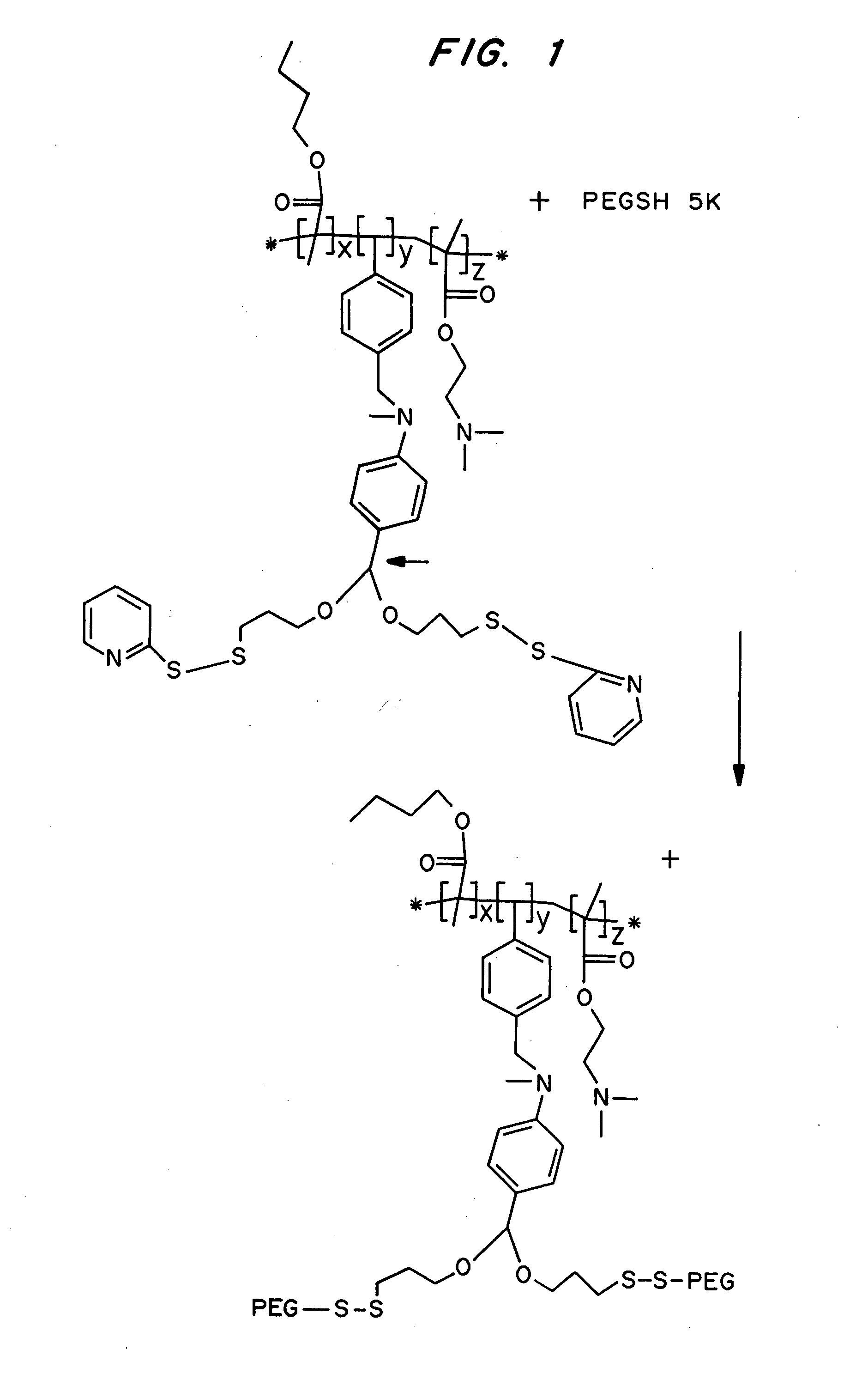
pH Mediated Disruption of Membranes using Acetal-PEG-Copolymer

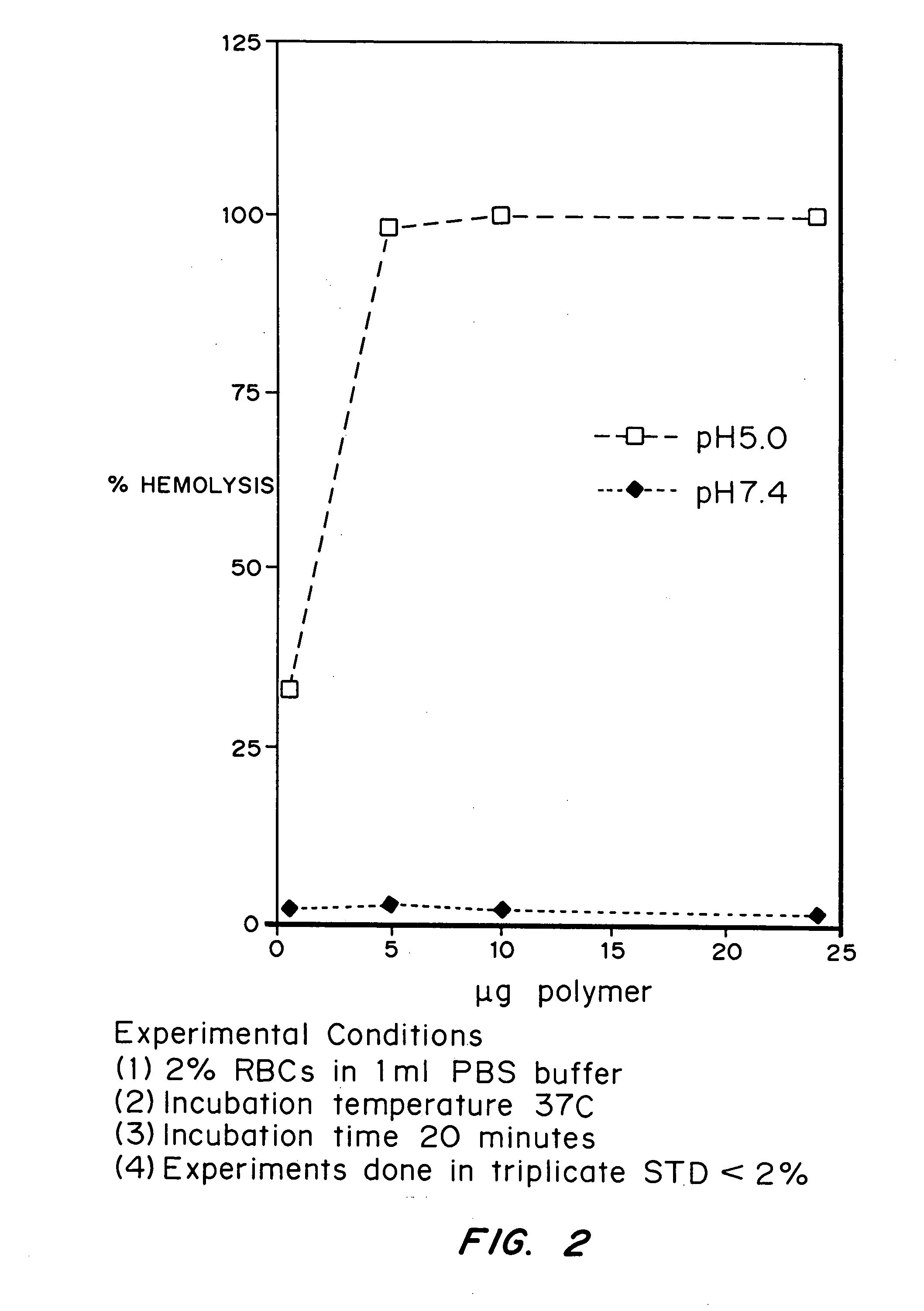
[0136]This example demonstrates that an acetal-PEG-copolymer can act as an endosomal releasing agent. This was determined by measuring the hemolytic activity of the above polymers at endosomal pH (5.5) and physiologic pH (7.4).

[0137]Synthesis of the polymeric conjugate is shown in FIG. 1.

[0138]Hemolysis Assay: Fresh human blood was isolated in EDTA containing vacutainers, washed three times with 150 mM NaCl, and resuspended at a concentration of 108 red blood cells / ml in PBS buffer (2% in 1 ml PBS) at either pH 5.5 or pH 7.4. The polymer was dissolved pH 10 buffered PBS. The appropriate volume of polymer solution was then added to the RBC solution and incubated for 20 minutes at 37° C. The cells were then centrifuged and the degree of hemolysis was determined by measuring absorbance of the supernatant at 541 nM. A 100% lysis was determined by lysing the red blood cells in deionized water. The controls were RBCS suspended ...

example 2

Synthesis of Compositions Containing a Terpolymer of Dimethylaminoethyl Methacrylate, Butyl Methacrylate and Styrene Benzaldehyde for pH Mediated Disruption of Membranes

[0140]A terpolymer of dimethylaminoethyl methacrylate (DMAEMA), butyl methacrylate (BMA) and styrene benzaldehyde was chosen for the membrane-disruptive backbone (see FIG. 4). Copolymers of BMA and DMAEMA are extremely effective membrane disruptive agents, a property that can be attributed to their cationic and hydrophobic components, leading to a surfactant-like character. C. Hansch, W. R. Glave, Mol. Pharmacol, 7:337 (1972).

[0141]The strategy used to synthesize the compositions is depicted in FIG. 5. The first step was the synthesis of a functionalized acetal monomer. This monomer was then copolymerized with DMAEMA and BMA using a free radical polymerization process. The resulting terpolymer was purified by precipitation in hexane and then PEGylated with thiol-terminated monofunctional or heterobifunctional PEGs. T...

example 3

Hydrolysis and Hemolysis Studies with Polymer E1

[0142]The hydrolysis of polymer E1 was measured at 37° C., in phosphate buffer, at either pH 5.4 or 7.4, by observing the change in U.V. absorbance at 340 nm. The experiments were done in triplicate and the standard deviation was under 5% for all samples.

[0143]The ability of polymer E1 to stimulate pH-induced membrane disruption was tested by assaying its ability to disrupt red blood cell membranes (RBCs) at pH 5.0 and pH 7.4 (FIG. 6B). One-hundred million RBCs in a 1 ml volume of phosphate buffer were used in each experiment. The incubation time was 20 minutes at 37° C. The experiments were done in triplicate and the standard deviation was under 5% for all samples. The protocol used to isolate and purify the red blood cells and quantitate hemolysis is described in N. Murthy, J. R. Robichaud, D. A. Tirrell, P. S. Stayton, A. S. Hoffman, J. Contr Rel, 61:137 (1999).

[0144]Only 0.5-5 μg / ml of the polymer E1 was required for efficient hemo...


PUM

PropertyMeasurementUnit
pHaaaaaaaaaa
pHaaaaaaaaaa
pHaaaaaaaaaa

Abstract

Compositions and methods for transport or release of therapeutic and diagnostic agents or metabolites or other analytes from cells, compartments within cells, or through cell layers or barriers are described. The compositions include a membrane barrier transport enhancing agent and are usually administered in combination with an enhancer and / or exposure to stimuli to effect disruption or altered permeability, transport or release. In a preferred embodiment, the compositions include compounds which disrupt endosomal membranes in response to the low pH in the endosomes but which are relatively inactive toward cell membranes (at physiologic pH, but can become active toward cell membranes if the environment is acidified below ca. pH 6.8), coupled directly or indirectly to a therapeutic or diagnostic agent. Other disruptive agents can also be used, responsive to stimuli and / or enhancers other than pH, such as light, electrical stimuli, electromagnetic stimuli, ultrasound, temperature, or combinations thereof. The compounds can be coupled by ionic, covalent or H bonds to an agent to be delivered or to a ligand which forms a complex with the agent to be delivered. Agents to be delivered can be therapeutic and / or diagnostic agents. Treatments which enhance delivery such as ultrasound, iontopheresis, and / or electrophereis can also be used with the disrupting agents.

Description

[0001]This application claims priority to U.S. Ser. No. 60 / 174,893 filed Jan. 7, 2000.STATEMENT REGARDING FEDERALLY FUNDED RESEARCH[0002]The U.S. government has certain rights in this invention by virtue of a National Institutes of Health grant, National Institutes General Medical Sciences grant GM 53771-02, 03 to 05.[0003]The present invention is in the field of two uses of acid-sensitive polymers: (1) intracellular release of agents from inside endosomes into the cytosol, and (2) lysis of cells and microorganisms from outside, for subsequent separation, recovery, and / or analysis of their contents. In the first use, the polymer is combined with other components such as therapeutic or diagnostic agents, targeting ligands, masking molecules. In the second use, the polymer is used as a free polymer, but may be combined with chemical or physical agents used to separate, recover, identify, assay, and / or label specific intracellular components.BACKGROUND OF THE INVENTION[0004]Specific, e...

Claims


Application Information

Patent Timeline
Patent Type & Authority Applications(United States)
IPC IPC(8): A61K38/16A61K38/00A61K31/70A61K31/7088A61K31/715C08F26/02C08F16/34C08F18/02C08F16/12A61P43/00A61P3/02A61P3/06A61K47/48
CPCA61K47/48169A61K47/48692A61K47/48238A61K47/56A61K47/62A61K47/6883A61P3/02A61P3/06A61P43/00
Inventor HOFFMAN, ALLAN S.STAYTON, PATRICK S.MURTHY, NIREN
Owner UNIV OF WASHINGTON
Learn More