
Subcutaneous access device and related methods

a subcutaneous access and access device technology, applied in the field of subcutaneous access devices, can solve the problems of affecting the operation efficiency of the peripheral vein, the fragile and limited peripheral vasculature of the patient, and the inability to locate the vein in the critically injured or ill patient, and achieve the effect of facilitating hands-free transillumination of the body portion

Active Publication Date: 2011-01-13
ENTROTECH INC
View PDF37 Cites 22 Cited by
  • Summary
  • Abstract
  • Description
  • Claims
  • Application Information

AI Technical Summary

Benefits of technology

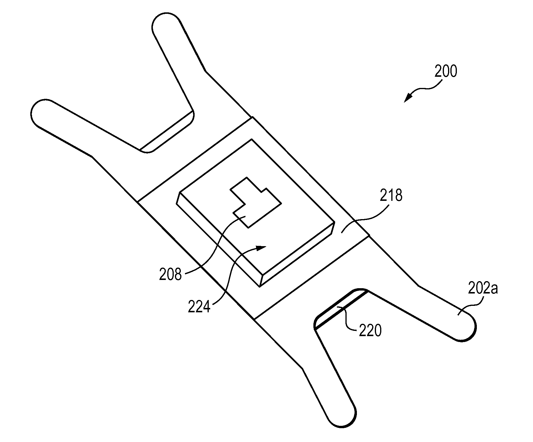
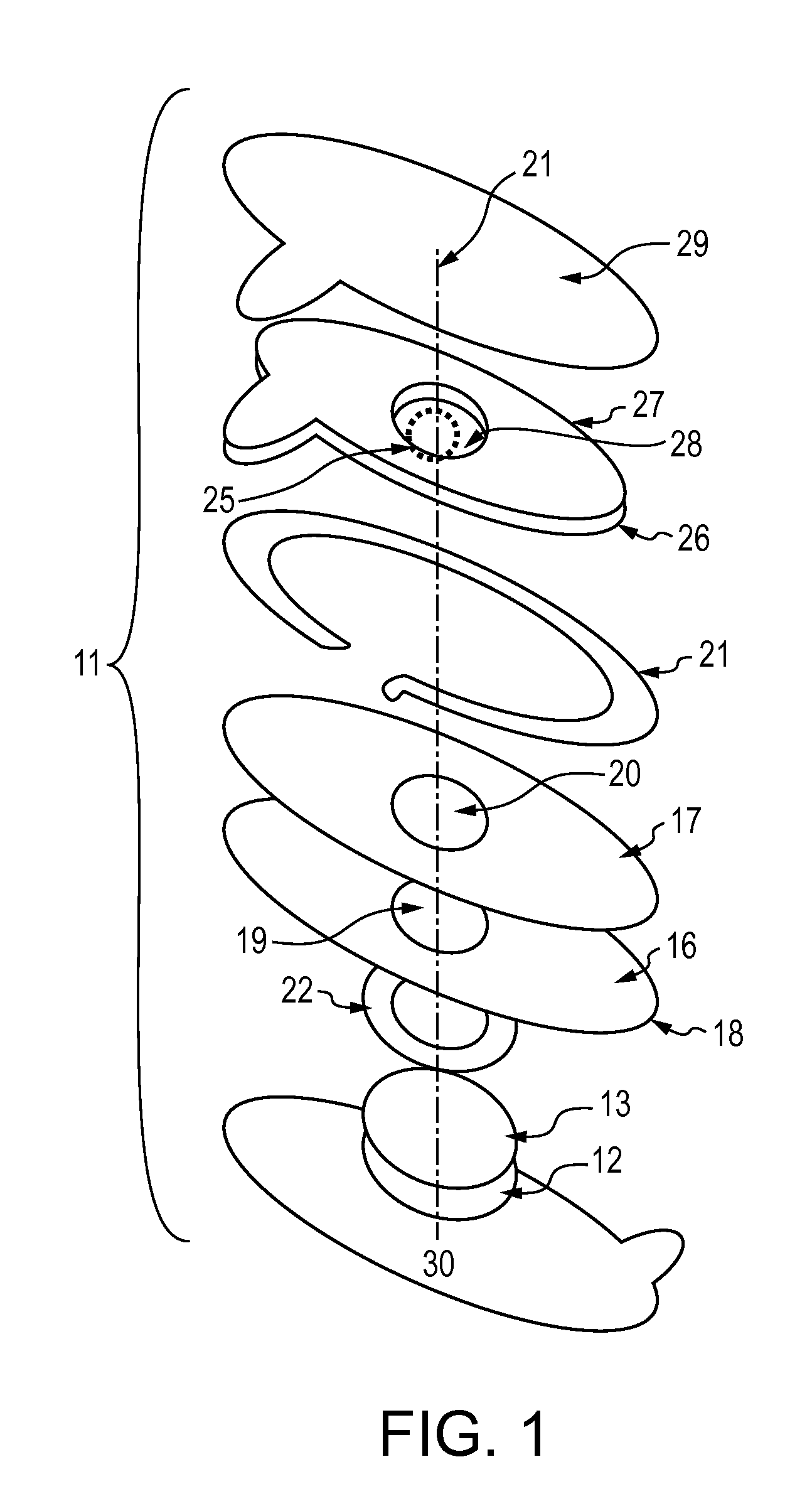
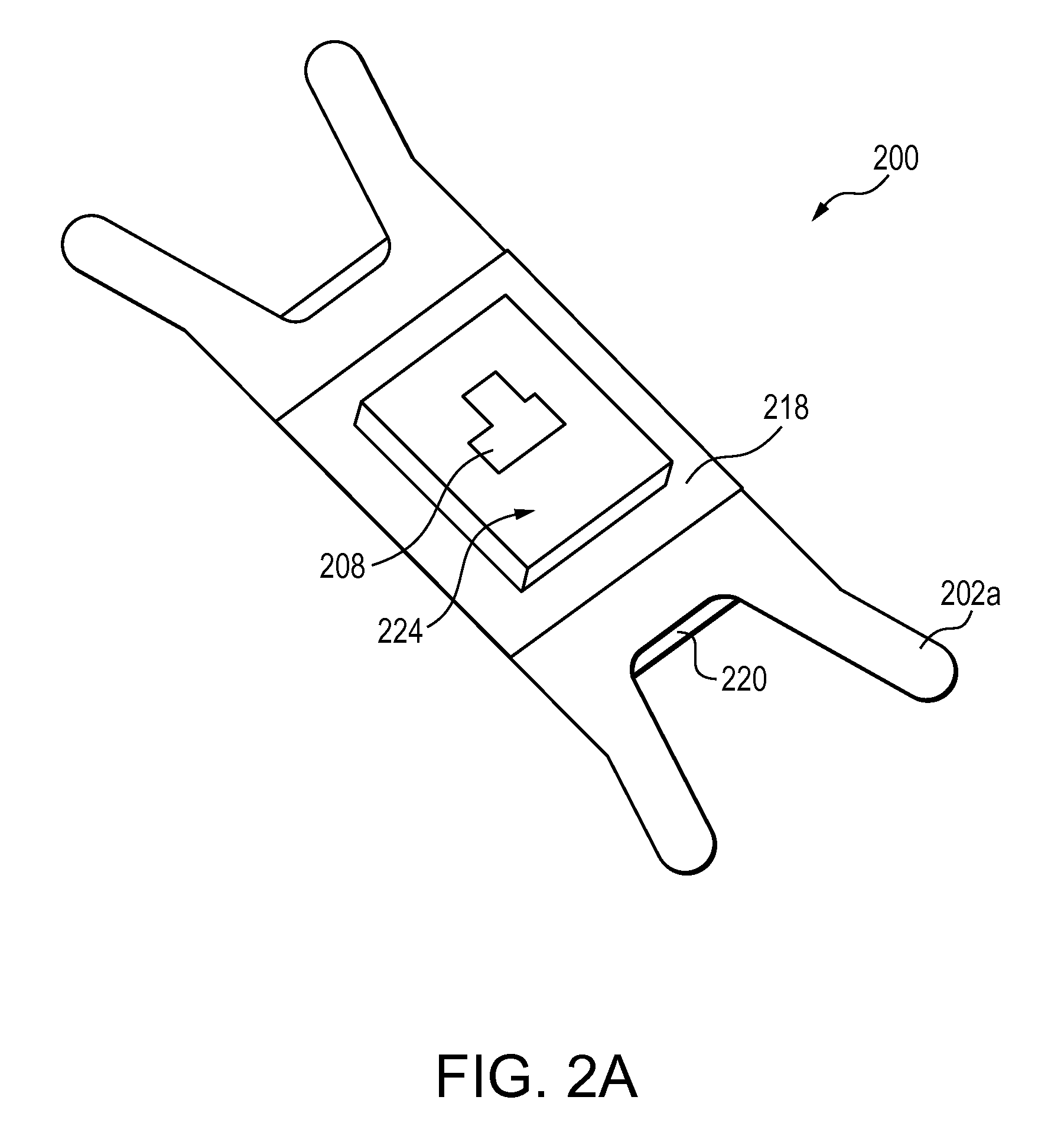
[0011]A subcutaneous access device of the invention comprises: a multi-layered structure capable of use in a medical imaging procedure; a light source for transillumination of a body portion of interest of a patient's body; and an attachment portion for attaching the device to the patient's body in a manner such that the body portion of interest is outwardly exposed on a side of the patient's body opposite the light source. The device facilitates hands-free transillumination of the body portion of interest. Kits for subcutaneous access comprise the subcutaneous access device and a light detector for detecting light emitted from the subcutaneous access device.

Problems solved by technology

For example, in emergencies, every second counts, and finding a vein in a critically injured or ill patient can be highly difficult and time-consuming when delivery of medications, fluids, and / or nutrients is most critical.
In oncology, frequent infusion therapies often make a patient's peripheral vasculature fragile and limited.
In the operating room, unsuccessful attempts at peripheral intravenous access delay many operations.
When peripheral access cannot be attained and a central venous catheter must be placed, a patient's risk for infection—particularly risky bacterial staph infections—goes up significantly.
In pediatrics, children pose a particular challenge for venipuncture due to their relatively small vessels and limited access points.
Often adding to these challenges are the fears of the child and the great anxiety of their parents.
When the patient is in the highly sensitive pediatric and neonatal intensive care unit, the situation can be even more challenging.
In the administration of medical care to a patient requiring subcutaneous access, especially in an emergency situation, such as that encountered by a physician, nurse, emergency medical technician (EMT), or other health care provider, or in the treatment of an accident victim at the scene of the accident, or in the treatment of wounded on a battlefield by a medic, environmental conditions under which medical care is administered may be adverse.
Further, during transport of a victim to a hospital or similar medical care facility, administration of medical procedures may be necessary, but difficult to perform, under poor lighting conditions or under other adverse conditions (e.g., those associated with torn clothing, bleeding, or other circumstances).
Shock may have caused veins of a victim to partially collapse, or the patient may have veins that are difficult to find (such as in the treatment of infants or obese persons), which further complicates procedures for accessing the veins.
Having subcutaneous fluid extracted (e.g., having blood drawn) or starting an intravenous access can be an uncomfortable, or even painful, experience for patients.
In addition to a fear of needles, many patients must unfortunately endure multiple needle sticks when a nurse or technician cannot easily find an appropriate access point.
Not surprisingly, even with adequate environmental conditions, finding a subcutaneous structure such as a vein is not always an easy task.
A number of factors can make it difficult to find a patient's veins, including: age, frequent (or lengthy) infusion therapies, multiple disease states, dehydration, hypertension, size and fragility of blood vessels, skin tone, and presence of edema or fatty tissue.
Despite advances in imaging, especially for visualization of subcutaneous structures in a patient's body, current systems tend to be cumbersome and inefficient.
For example, in known procedures for visualization of subcutaneous structures, proper support of a light source in order to accurately direct the light onto a body portion of interest may be an awkward procedure for the health care provider when treating a patient.
Moreover, current devices distinctly comprise light source(s) and support structure(s) that are essentially separate components that may be used together but are not intrinsically combined as an optimized device and as such have significant light losses and inefficiencies.

Method used


Image

  • Subcutaneous access device and related methods
  • Subcutaneous access device and related methods
  • Subcutaneous access device and related methods

Examples


Embodiment Construction

[0021]Subcutaneous access devices and methods of the invention facilitate rapid and convenient visualization of and easy and comfortable access to veins, arteries, and other subcutaneous structures within a patient's body. In contrast to conventional methods and apparatus for subcutaneous access, devices of the invention are advantageously portable and capable of efficient use in a variety of environments. Devices of the invention are capable of being simply and efficiently used when providing both routine and critical care in the administration of medical treatment to a patient. In exemplary embodiments, devices and methods of the invention are capably used in the field (e.g., proximate a battlefield) or in temporary battlefield installations (e.g., forward surgical units).

[0022]Devices of the invention are particularly useful in conjunction with and are an improvement to systems and methods described in U.S. Pat. No. 6,230,046 and U.S. Patent Publication No. 2007 / 0032721. Accordin...


PUM


Abstract

A subcutaneous access device of the invention comprises: a multi-layered structure capable of use in a medical imaging procedure; a light source for transillumination of a body portion of interest of a patient's body; and an attachment portion for attaching the device to the patient's body in a manner such that the body portion of interest is outwardly exposed on a side of the patient's body opposite the light source. The device facilitates hands-free transillumination of the body portion of interest. Kits for subcutaneous access comprise the subcutaneous access device and a light detector for detecting light emitted from the subcutaneous access device.

Description

BACKGROUND OF THE INVENTION[0001]Many methods and apparatus for accessing subcutaneous structures (e.g., vessels, such as veins and arteries, or other structures) are known. One common need for subcutaneous access involves intravenous insertion or extraction of fluids, medications, or the like when administering medical treatment to a patient. Subcutaneous access may be needed in, for example, the following medical facilities and departments: emergency, labor and delivery, laboratory, oncology, operating room, intensive care unit, pediatrics, and pediatric and neonatal intensive care unit.[0002]For example, in emergencies, every second counts, and finding a vein in a critically injured or ill patient can be highly difficult and time-consuming when delivery of medications, fluids, and / or nutrients is most critical. In labor and delivery, an intravenous access device is often immediately placed in a patient during the admitting process for hydration or to introduce medication. Pregnan...

Claims


Application Information

Patent Timeline
Patent Type & Authority Applications(United States)
IPC IPC(8): A61B6/00
CPCA61B5/0086A61B5/6833A61B5/6824A61B5/489
Inventor MCGUIRE, JR., JAMES E.CALLARD, DAVID MELANCTHON
Owner ENTROTECH INC
Learn More