X-ray imaging method and x-ray imaging system

Active Publication Date: 2011-03-31
SIEMENS HEATHCARE GMBH
View PDF14 Cites 7 Cited by
  • Summary
  • Abstract
  • Description
  • Claims
  • Application Information

AI Technical Summary

Benefits of technology

[0007]The object of the present invention is to provide a slot-scan method which makes do without mechanically-movable collimators and makes a simple and effortless recording of an x-ray image possible. Furthermore an object of the invention is to provide an x-ray device suitable for carrying out the method.
[0014]The inventive method thus replaces known slot scanning methods and is able to be carried out more quickly and more easily through the sequential activation of the x-ray emitters and their already pre-collimated x-ray emitters. The recording time is greatly reduced since this is no longer dictated by the mechanical movement of the collimator but only by the tube power of the x-ray emitter and by the read-out time of the x-ray detector. In addition scattered radiation can be almost completely avoided by the inventive method and thus high-quality x-ray images can be created.
[0019]To carry out the inventive method a medical x-ray imaging system with an x-ray source and an x-ray detector is provided, with the x-ray source featuring a plurality of x-ray emitters arranged next to one another. In this case the x-ray emitters can be formed in each case by a field emission gun with a field emission cathode. Such field emission guns can be made especially small and light. In accordance with a further embodiment of the invention the field emission cathodes are formed on the basis of carbon nanotubes (so called. CNT cathodes). These types of material exhibit an especially good emission characteristic but are also stable with high currents and can also be manufactured especially small. In an advantageous manner the field emission guns are arranged as a linear array. This array is disposed so that a simple assignment to the respective, especially row-type, part areas of the x-ray detector is possible.

Problems solved by technology
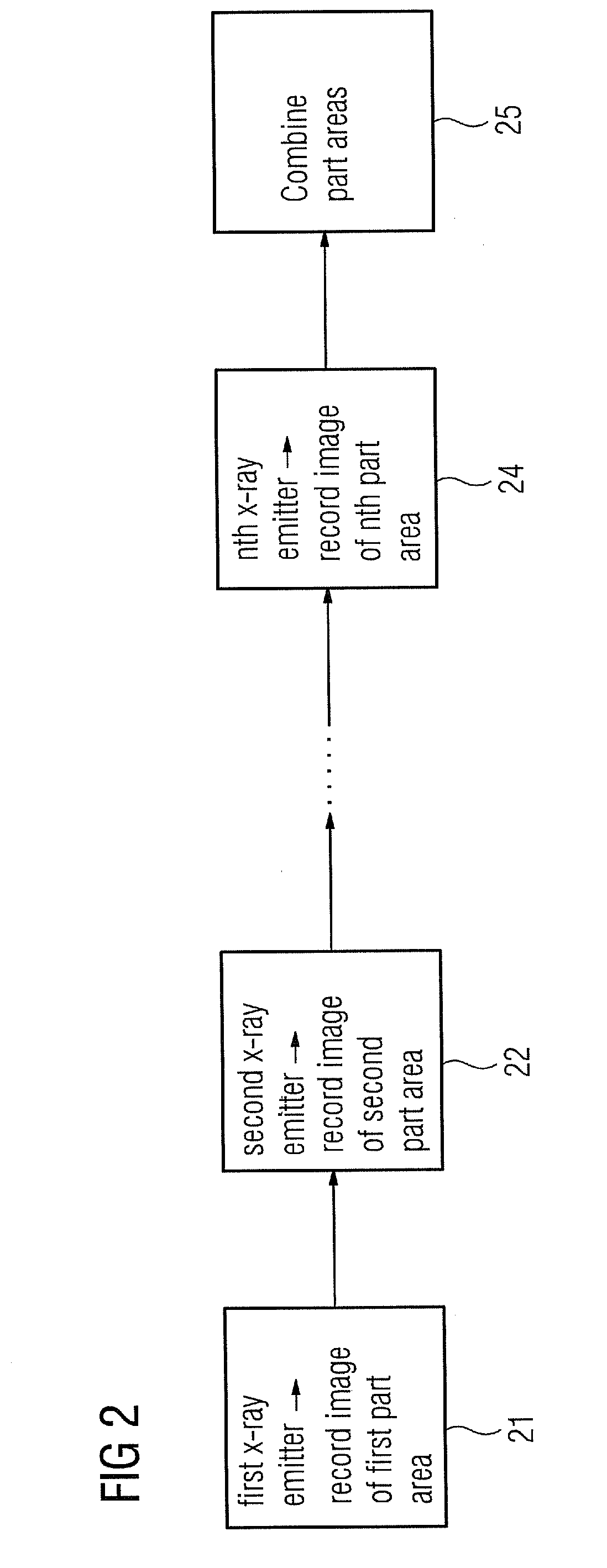
A problem with x-ray imaging is that the image quality is adversely affected by what is known as radiation scatter.
Anti-scatter grids have the problem however that on the one hand they still let through part of the scattered radiation and on the other hand they also absorb a significant part of the direct x-ray radiation (up to 50%).
Despite greatly improving radiation scatter, slot scanning systems have as yet not become established.
However this system still needs a mechanically-movable tube-side collimator.

Method used


Image

  • X-ray imaging method and x-ray imaging system
  • X-ray imaging method and x-ray imaging system
  • X-ray imaging method and x-ray imaging system

Examples


Embodiment Construction

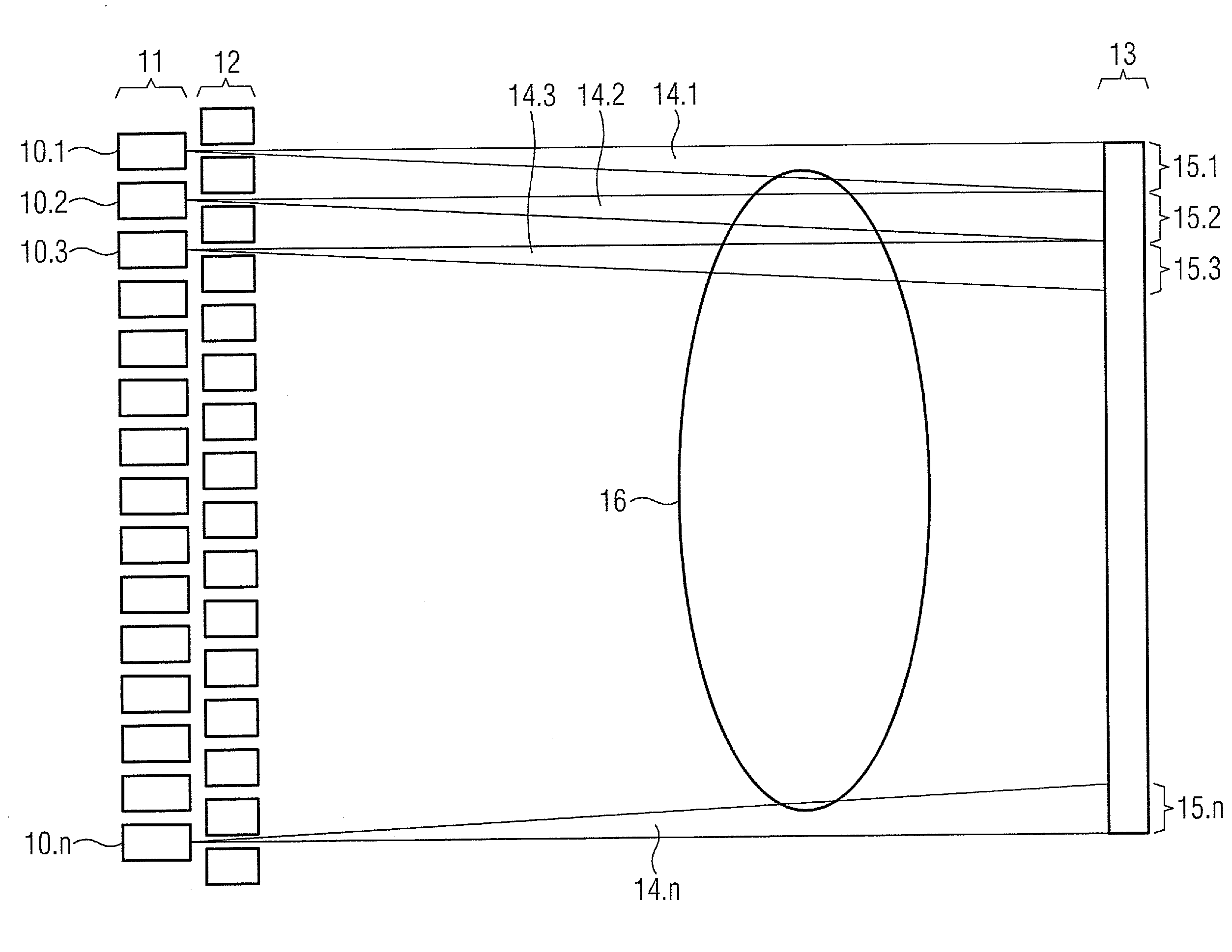
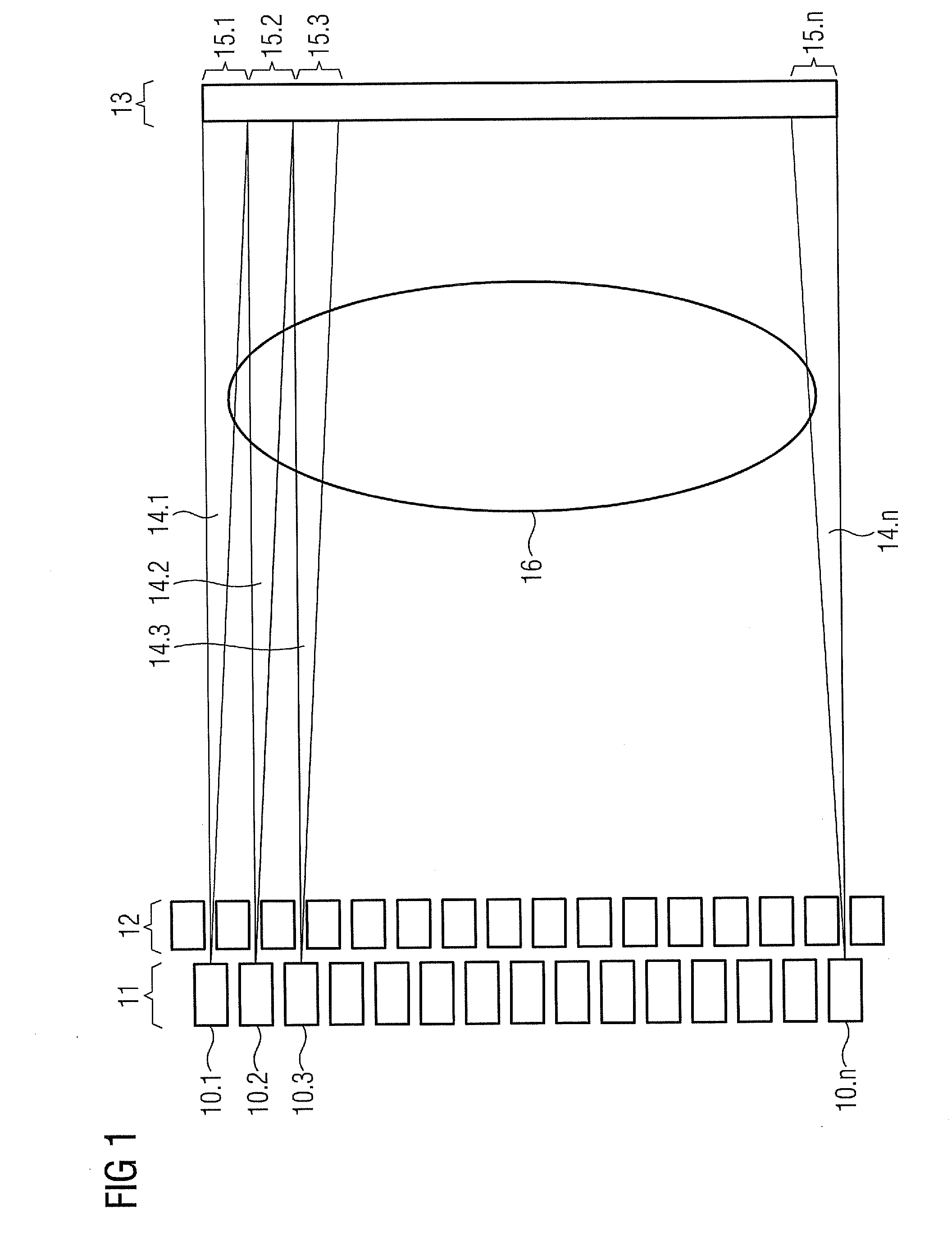
[0024]FIG. 1 shows an inventive x-ray imaging system with an x-ray source 11 in the form of a plurality of x-ray emitters 10, especially field emission guns, and a digital x-ray detector 13, with part areas 15 of the x-ray detector assigned in each case to the x-ray emitters 10. The x-ray emitters 10 are arranged alongside one another in a linear array. Each x-ray emitter 10 is embodied to emit an x-ray beam 14, with the x-ray beams 14 being formed by a collimator 12 into slots, so that each x-ray beam 14 strikes a part area of 15 of the x-ray detector. The collimation by means of the collimator can be set when the x-ray imaging system is commissioned or the device can already have a fixed setting on delivery. The digital x-ray detector especially involves a known digital solid-state detector based on direct or indirect conversion of x-rays into an electrical charge.

[0025]A first x-ray beam 14.1 created by a first x-ray emitter 10.1 is formed in this case by the collimator 12 or by ...


PUM


Abstract

A method for recording an x-ray image with an x-ray imaging system having an x-ray source and an x-ray detector is provided. The x-ray source has a plurality of x-ray emitters arranged alongside one another. Each x-ray emitter is assigned to a part area of the x-ray detector. The x-ray beam generated by the x-ray emitter is collimated onto the assigned part area. A first x-ray emitter is activated for emitting a first x-ray beam onto a first part area and image data of the first part area is read out. Subsequently a further x-ray emitter for emitting a further x-ray beam onto a further part area of is activated and image data of the further part area is read out. The steps are repeated until image data for all part areas of the x-ray detector is read out. The read out image data is combined into an x-ray image.

Description

CROSS REFERENCE TO RELATED APPLICATIONS[0001]This application claims priority of German application No. 10 2009 043 423.2 filed Sep. 29, 2009, which is incorporated by reference herein in its entirety.FIELD OF THE INVENTION[0002]The invention relates to a method for recording an x-ray image with an x-ray imaging system as well as to a device for carrying out such a method.BACKGROUND OF THE INVENTION[0003]X-ray radiography systems are used for diagnostic imaging, whereby they provide image data of a region of a patient's body to be examined in order to make possible or to simplify a medical diagnosis. The image data is obtained with the aid of an x-ray source and an x-ray-sensitive x-ray detector. A modern digital x-ray system is described in U.S. Pat. No. 6,733,176 for example.[0004]A problem with x-ray imaging is that the image quality is adversely affected by what is known as radiation scatter. Radiation scatter occurs when x-ray radiation not only strikes the x-ray detector on a ...

Claims


Application Information

Patent Timeline
IPC IPC(8): G01N23/04
CPCA61B6/06A61B6/5282G21K1/025H01J2235/068H01J35/065H01J2235/062G21K2207/00
Inventor BOESE, JANMASCHKE, MICHAEL
Owner SIEMENS HEATHCARE GMBH
Why Eureka
Try Eureka