Tissue closure device and method

a tissue closure and tissue technology, applied in the field of tissue closure devices and methods, can solve the problems of difficult and complicated closure methods such as suturing, difficult and complicated application of sutures with remotely operated thoracoscopic instruments, and difficult closing of small openings, so as to prevent the driving of anchors

Active Publication Date: 2012-01-26
MICRO INTERVENTIONAL DEVICES
View PDF12 Cites 34 Cited by
  • Summary
  • Abstract
  • Description
  • Claims
  • Application Information

AI Technical Summary

Benefits of technology

[0022]The hooked projection may be configured to maintain engagement between the band and the anchor by preventing the band from moving off the proximal end of the anchor.
[0031]The closure element may be prevented from extending within the ring-shaped circumference by one or more tubes.
[0057]The hooked projection may be configured to maintain engagement between the band and the anchor by preventing the band from moving off the proximal end of the anchor.
[0066]The key plate may prevent driving of the anchors when the at least one key member is engaged with the key plate.
[0067]The at least one key member includes a plurality of key members each being independently movable by a respective contact element. The key plate may prevent driving of the anchors if any one of the key members is engaged with the key plate.
[0069]The closure element may be prevented from extending within the ring-shaped circumference when the anchors are maintained in the first configuration.

Problems solved by technology

However, these small openings may be difficult to close, especially where the closure location is inside the patient's body.
Since these exemplary thoracoscopic procedures involve accessing the *patient's thorax through a small intercostal aperture through the patient's skin and other underlying tissues (e.g., fat and / or fascia), closure methods such as suturing are more complicated than with non-thoracoscopic surgical procedures.
In particular, applying sutures with remotely operated thoracoscopic instruments is more difficult and complicated than directly manipulating a suture needle by hand at the surgical site.
This difficulty can result in defective closures and / or closures that require more time than necessary.
Defective closures may expose the patient to increased risk of complications such as internal bleeding and / or infection.
Even where defective closures are recognized and addressed prior to completion of the surgical procedure, the correction of defective closures increases the time required to effect the closure and may expose the tissue to additional trauma.

Method used


Image

  • Tissue closure device and method
  • Tissue closure device and method
  • Tissue closure device and method

Examples


Embodiment Construction

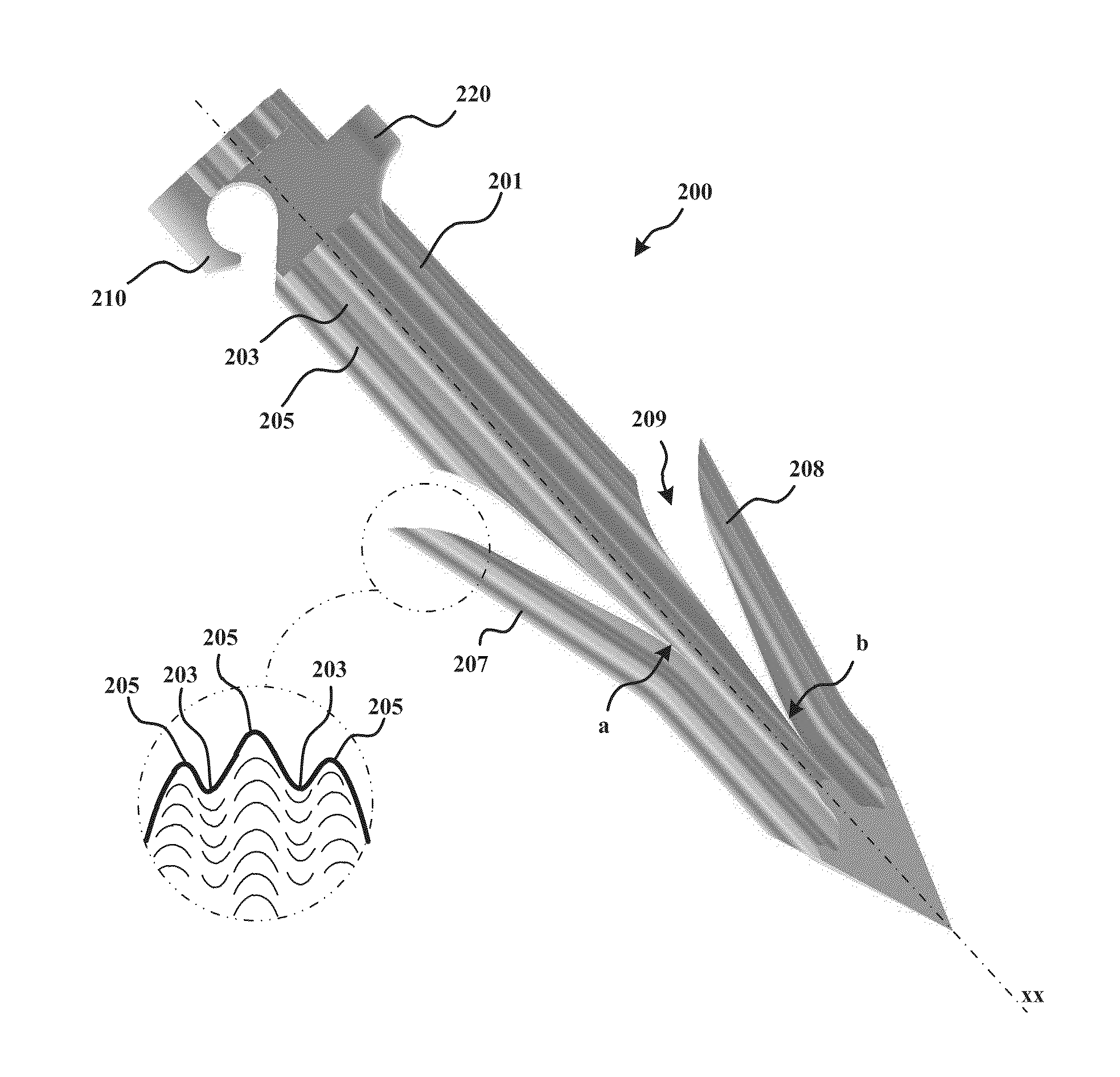
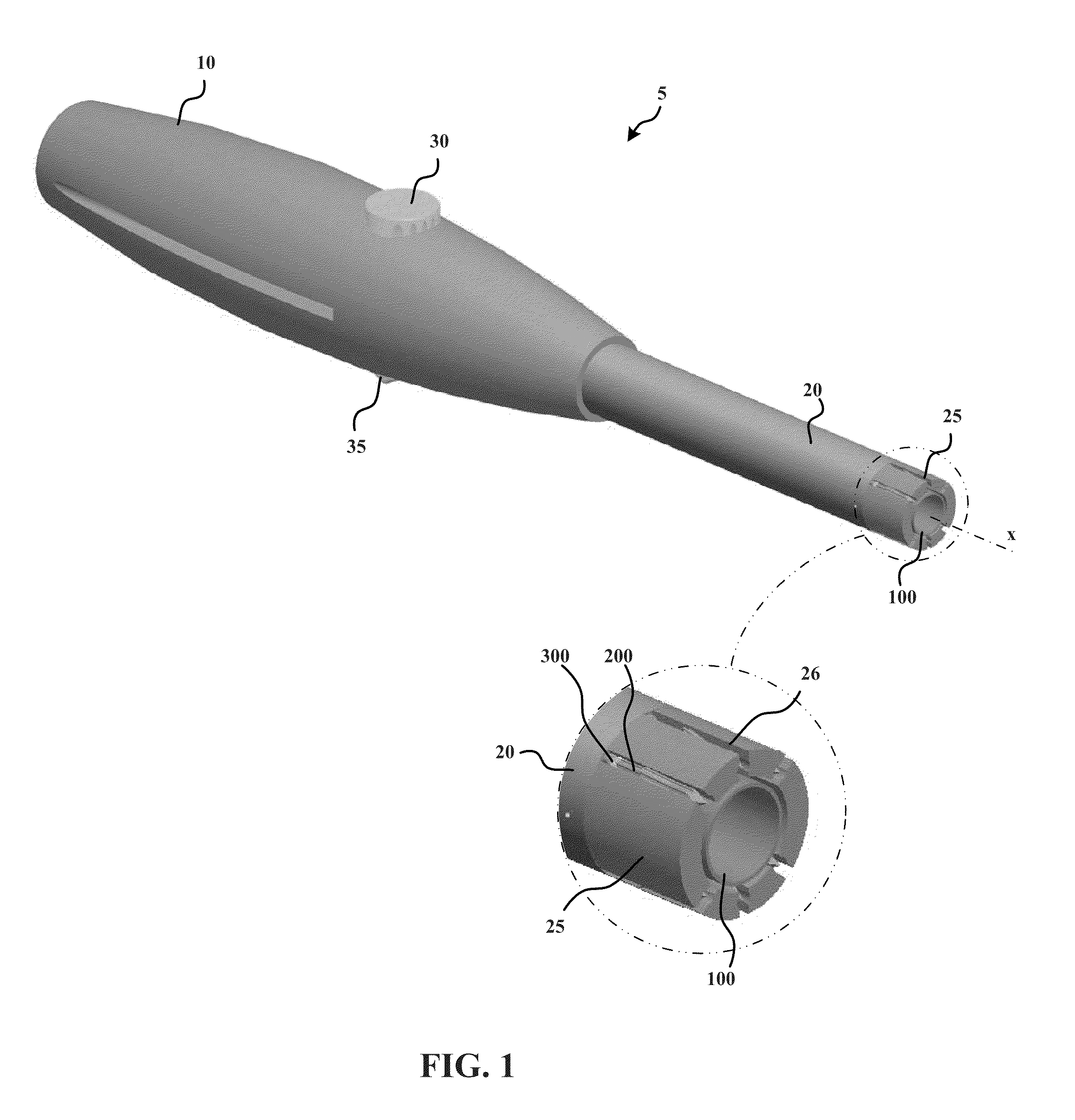
[0142]As set forth in greater detail below, example embodiments of the present invention allow for the reliable and effective closure of an opening in tissue (e.g., a pericardial window) that limits the possibility of human error, e.g., by eliminating the need for suturing. In some examples, a surgical device anchors a plurality of anchors, which are connected to each other by one or more elastic closure elements, into the tissue. The anchors are driven into the tissue in a spaced-apart configuration in which the elastic closure elements are tensioned between the anchors. The anchors are held in the spaced-apart arrangement while a surgical procedure is performed through a tissue opening formed between the anchored locations of the anchors. In order to close the opening, the device simply releases the anchors from the spaced-apart arrangement such that the tensioned elastic closure elements draw the anchors, as well as the tissue in which the anchors are anchored, toward the tissue ...


PUM


Abstract

A device having a plurality of anchors configured to be driven into a tissue, and a closure element extending between the anchors and configured to urge the anchors from a first configuration in which the anchors are a first distance from each other, toward a second configuration in which the anchors are a second distance from each other, the second distance being less than the first distance, wherein the device is configured to maintain the anchors in the first configuration during a surgical intervention and to subsequently allow the anchors to be moved by the closure element toward the second configuration.

Description

CROSS-REFERENCE TO RELATED APPLICATIONS[0001]This application claims the benefit of U.S. Provisional Patent Application Ser. No. 61 / 296,868, filed on Jan. 20, 2010, which is hereby incorporated herein in its entirety by reference thereto.[0002]Further, each of the following is hereby incorporated in its entirety by reference thereto: U.S. patent application Ser. No. ______, Attorney Docket No. 14895 / 3, filed on Jan. 20, 2011, U.S. patent application Ser. No. ______, Attorney Docket No. 14895 / 4, filed on Jan. 20, 2011; and U.S. patent application Ser. No. ______, Attorney Docket No. 14895 / 5, filed on Jan. 20, 2011.FIELD OF THE INVENTION[0003]The present invention relates to a tissue closure device and method.BACKGROUND INFORMATION[0004]Some surgical interventions involve piercing or cutting a hole into a tissue wall. For example, thoracoscopic procedures typically involve piercing one or more tissue walls with a trocar or other sharp device and insertion of a cannula to maintain an o...

Claims


Application Information

Patent Timeline
Patent Type & Authority Applications(United States)
IPC IPC(8): A61B17/03
CPCA61B17/0057A61B17/0401A61B2017/00575A61B2017/00579A61B2017/00632A61B2017/0408A61B2017/0648A61B2017/0437A61B2017/0464A61B17/10A61B2017/0647A61B2017/0427A61B2017/0435A61B2017/0412A61B17/0487A61B2017/0414A61B2017/0488A61B2017/081
Inventor WHITMAN, MICHAEL P.DATCUK, PETER
Owner MICRO INTERVENTIONAL DEVICES
Why Eureka
Try Eureka