
Image segmentation

Active Publication Date: 2012-02-02
TOSHIBA MEDICAL SYST CORP
View PDF0 Cites 83 Cited by
  • Summary
  • Abstract
  • Description
  • Claims
  • Application Information

AI Technical Summary

Problems solved by technology

The overall goal of an organ atlas is to be as typical as possible to an anatomical category, and this poses a challenge to medical image analysis because of underlying variability in anatomy.

Method used


Image

  • Image segmentation
  • Image segmentation
  • Image segmentation

Examples


example

[0085]An example procedure for selecting the best set of atlas data sets to use for multi-atlas based segmentation is now described.

[0086]Cardiac CT angiography (CCTA) data sets from 21 patients were used to construct the training set. The data sets represent two distinct categories of heart anatomy: patients with small cardiothoracic ratio (CTR), and patients with large cardiothoracic ratios. Data sets 9, 12, and 16-21 were identified as being from patients with large CTRs. Whole heart segmentations, represented as masks containing the heart and all its sub-anatomies, were collected by a clinically qualified individual on each of these data sets. Each data set in turn is used as an atlas to segment the heart in all the other data sets. The segmentation is performed using multi-scale rigid registration to align the volumes based on 3-D translation, anisotropic scaling and rotation, followed by multi-scale non-rigid registration, which uses mutual information to find the best deforma...


PUM


Abstract

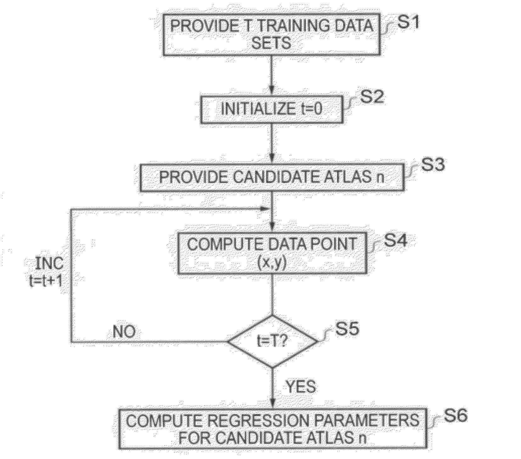
According to one embodiment there is provided a method of selecting a plurality of M atlases from among a larger group of N candidate atlases to form a multi-atlas data set to be used for computer automated segmentation of novel image data sets to mark objects of interest therein. A set of candidate atlases is used containing a reference image data set and segmentation data. Each of the candidate atlases is segmented against the others in a leave-one-out strategy, in which the candidate atlases are used as training data for each other. For each candidate atlas in turn, the following is carried out: registering; segmenting; computing an overlap; computing a value of the similarity measure for each of the registrations; and obtaining a set of regression parameters by performing a regression with the similarity measure being the independent variable and the overlap being the dependent variable. The M atlases are then selected from among all the N candidate atlases to form the multi-atlas data set, the M atlases being those atlases determined to collectively provide the highest aggregate overlap over all the training data image sets.

Description

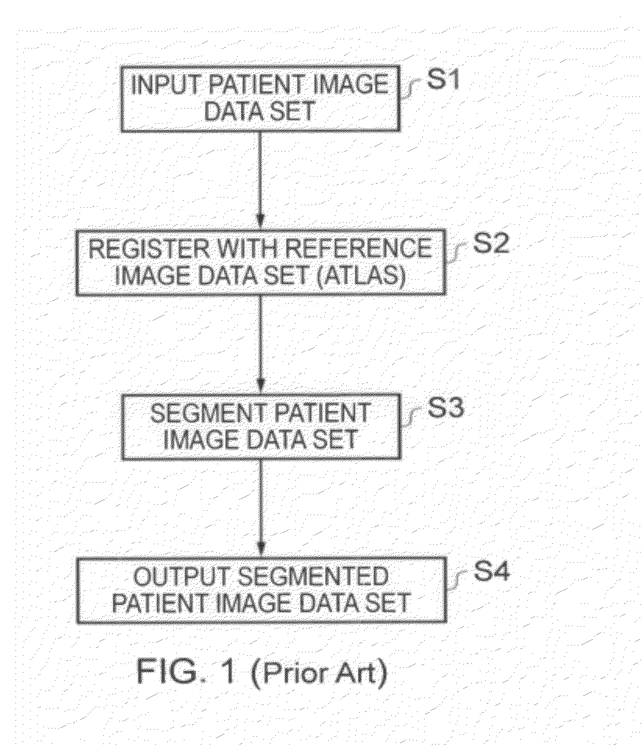
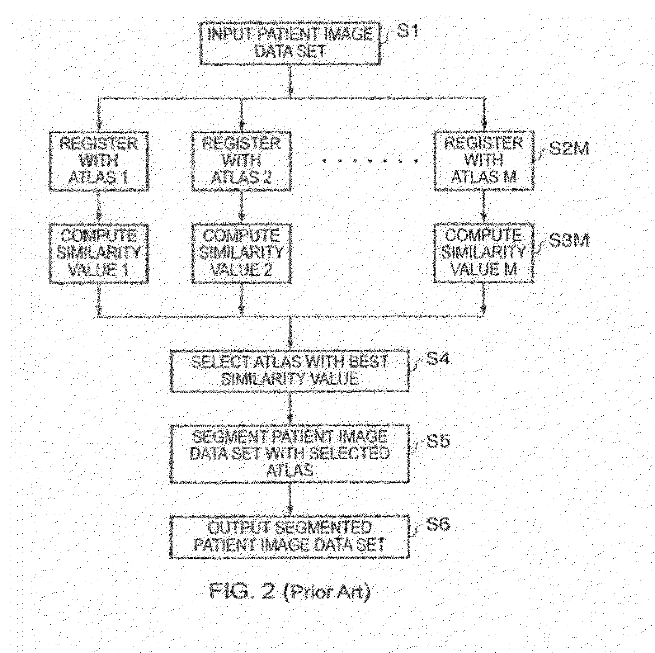
BACKGROUND OF THE INVENTION[0001]Embodiments described herein relate generally to segmentation of images, such as three-dimensional (3D) medical images.[0002]The method by which objects represented in images are labeled is referred to as segmentation.[0003]Objects may be manually labeled by an expert, and this is considered to be ‘the gold standard’ in view of the sophistication of an expert in recognizing objects, such as a clinician identifying organs and their sub-structure in two-dimensional (2D) or 3D X-ray images.[0004]Automated image processing procedures carried out by computers are widely available to perform segmentation in a host of different applications.[0005]FIG. 1 is a flow chart showing a basic segmentation process.[0006]In Step S1, a novel image data set, referred to in the figure as a patient image data set, is provided to a computer for segmentation.[0007]In Step S2, a registration is carried out to align the novel image data set with a reference image data set wh...

Claims


Application Information

Patent Timeline
IPC IPC(8): G06K9/34
CPCG06T7/0081G06T2207/30004G06T2207/20128G06T7/11Y10S128/922Y10S128/923Y10S128/924
Inventor AKINYEMI, AKINOLAPOOLE, IANPLAKAS, COSTASPIPER, JIM
Owner TOSHIBA MEDICAL SYST CORP
Learn More