Method and System for Liver Lesion Detection

a technology for liver lesions and detection methods, applied in image analysis, image enhancement, instruments, etc., can solve the problems of time-consuming and laborious manual finding of liver lesions

Active Publication Date: 2012-03-22
SIEMENS HEALTHCARE GMBH
View PDF3 Cites 41 Cited by
  • Summary
  • Abstract
  • Description
  • Claims
  • Application Information

AI Technical Summary

Benefits of technology

[0004]The present invention provides a method and system for automatically detecting and segmenting liver lesions. In particular, embodiments of the present invention utilize a robust and effective learning based method for automatic detection of liver lesions from computed tomography (CT) data. Embodiments of the present invention can automatically detect hypodense and hyperdense lesions from a single 3D CT image of any contrast phase.

Problems solved by technology

However, manually finding these lesions is tedious, time consuming, and highly dependent on the observer's experiences.

Method used


Image

  • Method and System for Liver Lesion Detection
  • Method and System for Liver Lesion Detection
  • Method and System for Liver Lesion Detection

Examples


Embodiment Construction

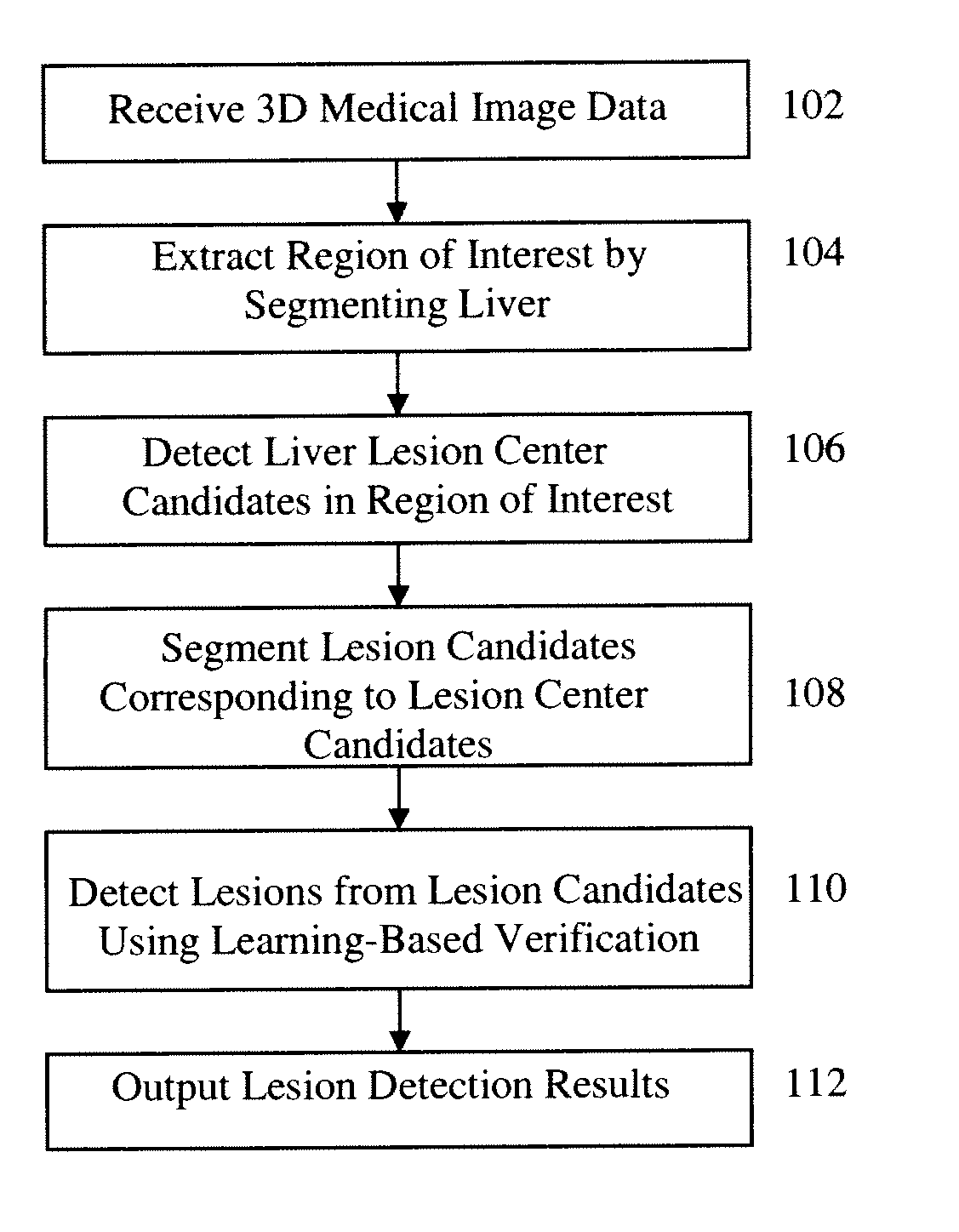
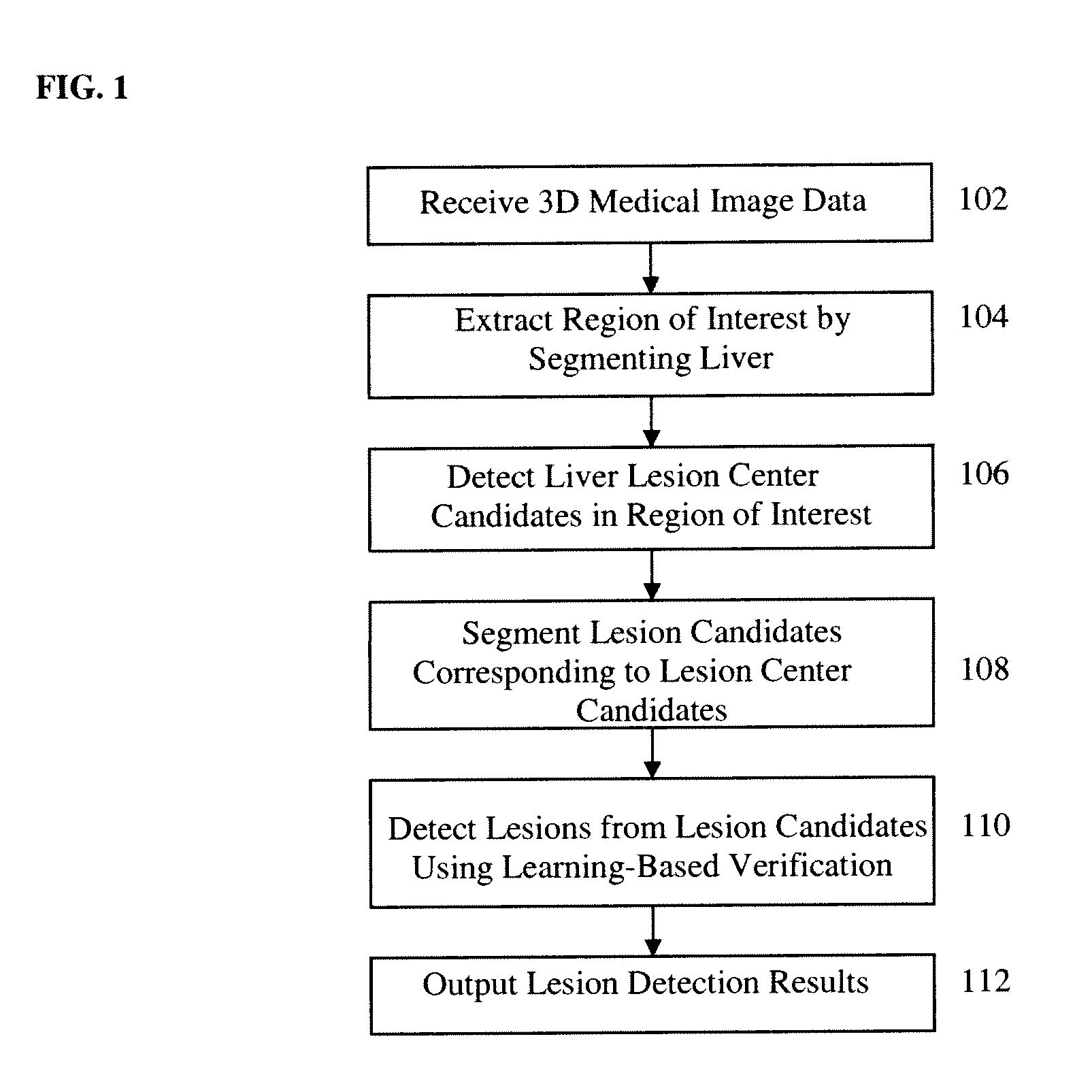
[0024]The present invention is directed to a method and system for automatically detecting liver lesions in medical image data. Embodiments of the present invention are described herein to give a visual understanding of the liver lesion detection method. A digital image is often composed of digital representations of one or more objects (or shapes). The digital representation of an object is often described herein in terms of identifying and manipulating the objects. Such manipulations are virtual manipulations accomplished in the memory or other circuitry / hardware of a computer system. Accordingly, it is to be understood that embodiments of the present invention may be performed within a computer system using data stored within the computer system.

[0025]Tumor staging and follow-up examinations account for a large part of routine work in radiology. Cancer patients regularly undergo a computed tomography (CT) examination in intervals of several weeks or months to monitor the patient ...


PUM


Abstract

A method and system for automatically detecting liver lesions in medical image data, such as 3D CT images, is disclosed. A liver region is segmented in a 3D image. Liver lesion center candidates are detected in the segmented liver region. Lesion candidates are segmented corresponding to the liver lesion center candidates, and lesions are detected from the segmented lesion candidates using learning based verification.

Description

[0001]This application claims the benefit of U.S. Provisional Application No. 61 / 385,221, filed Sep. 22, 2010, the disclosure of which is herein incorporated by reference.BACKGROUND OF THE INVENTION[0002]The present invention relates to detection of liver lesions, and more particularly, to database-guided liver lesion detection and segmentation in medical images.[0003]Detection of abnormal hepatic masses is important to liver disease diagnosis, treatment planning and follow-up monitoring. As a significant part of clinical practice in radiology, liver tumors are usually examined and tracked every several weeks or months to assess the cancer staging and therapy response based on 3D Computed Tomography (CT) data. However, manually finding these lesions is tedious, time consuming, and highly dependent on the observer's experiences. Hence, a system of automatic lesion detection and measurement is desirable.BRIEF SUMMARY OF THE INVENTION[0004]The present invention provides a method and sy...

Claims


Application Information

Patent Timeline
Patent Type & Authority Applications(United States)
IPC IPC(8): G06K9/00
CPCG06T7/0016G06T7/401G06T2207/10072G06T2207/30056G06T2207/20041G06T2207/20081G06T2207/20104G06T2207/20012G06T7/41
Inventor LIU, DAVIDWU, DIJIAZHOU, SHAOHUA KEVINCOSTA, MARIA JIMENASUEHLING, MICHAELTIETJEN, CHRISTIAN
Owner SIEMENS HEALTHCARE GMBH
Why Eureka
Try Eureka