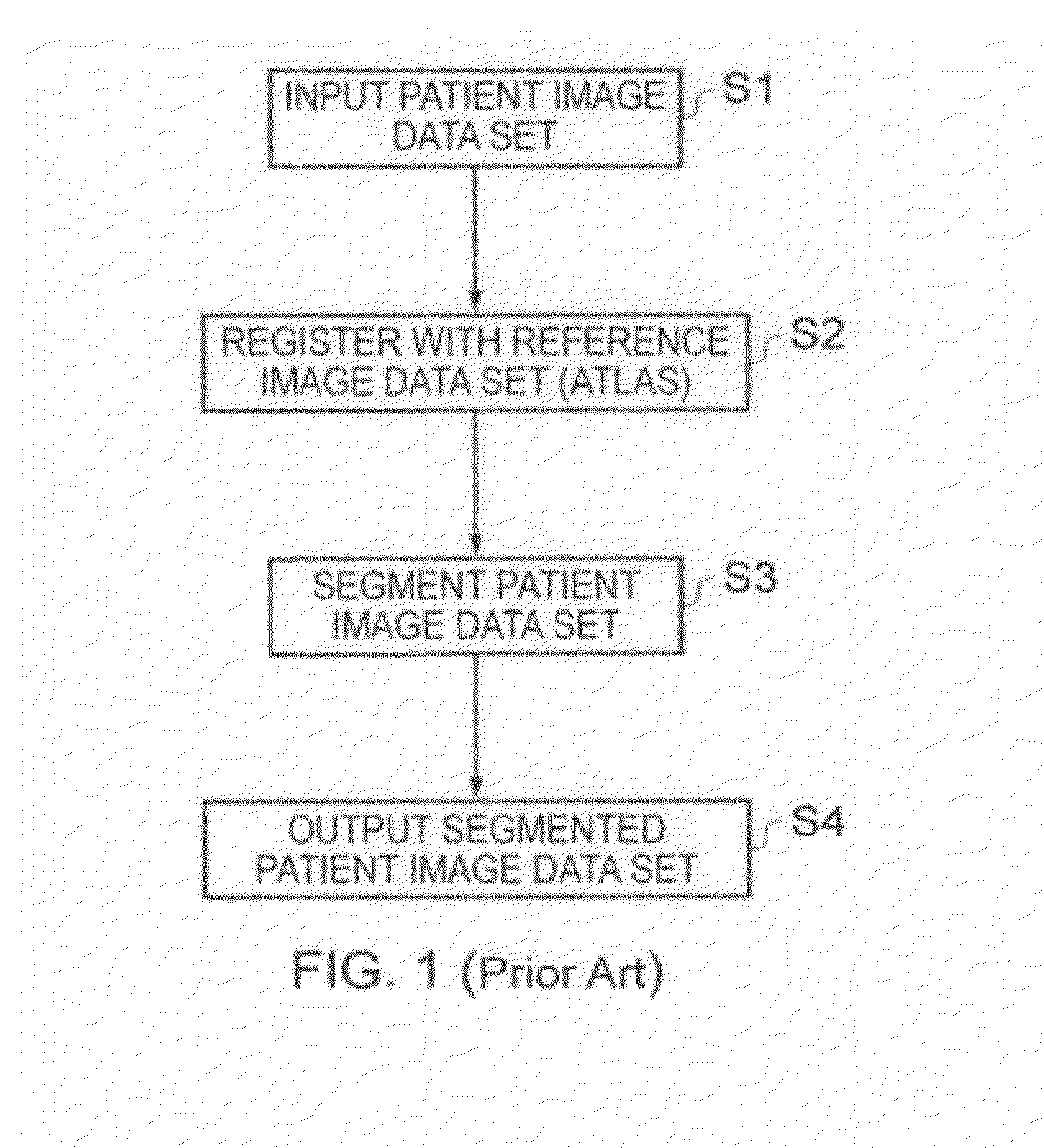
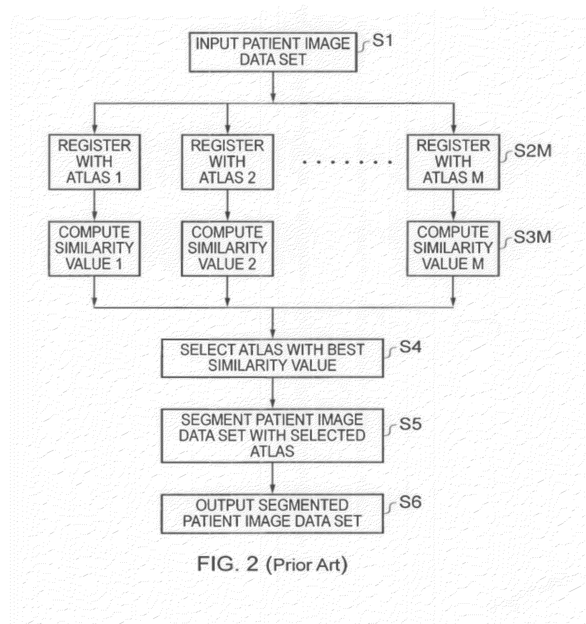
Image segmentation

US20120177263A1Active Publication Date: 2012-07-12TOSHIBA MEDICAL SYST CORP

Patent Information

Authority / Receiving Office
US Β· United States
Patent Type
Applications(United States)
Current Assignee / Owner
TOSHIBA MEDICAL SYST CORP
Publication Date
2012-07-12

Smart Images

  • Figure 1
    Figure 1
  • Figure 2
    Figure 2
  • Figure 3
    Figure 3
Patent Text Reader

Abstract

According to one embodiment there is provided a method of selecting a plurality of M atlases from among a larger group of N candidate atlases to form a multi-atlas data set to be used for computer automated segmentation of novel image data sets to mark objects of interest therein. A set of candidate atlases is used containing a reference image data set and segmentation data. Each of the candidate atlases is segmented against the others in a leave-one-out strategy, in which the candidate atlases are used as training data for each other. For each candidate atlas in turn, the following is carried out: registering; segmenting; computing an overlap; computing a value of the similarity measure for each of the registrations; and obtaining a set of regression parameters by performing a regression with the similarity measure being the independent variable and the overlap being the dependent variable.
Need to check novelty before this filing date? Find Prior Art

Description

CROSS-REFERENCE TO RELATED APPLICATION

[0001] This application is a divisional of U.S. application Ser. No. 12 / 847,372, filed Jul. 30, 2010.BACKGROUND OF THE INVENTION

[0002] Embodiments described herein relate generally to segmentation of images, such as three-dimensional (3D) medical images.

[0003] The method by which objects represented in images are labeled is referred to as segmentation.

[0004] Objects may be manually labeled by an expert, and this is considered to be β€˜the gold standard’ in view of the sophistication of an expert in recognizing objects, such as a clinician identifying organs and their sub-structure in two-dimensional (2D) or 3D X-ray images.

[0005] Automated image processing procedures carried out by computers are widely available to perform segmentation in a host of different applications.

[0006] FIG. 1 is a flow chart showing a basic segmentation process.

[0007] In Step S1, a novel image data set, referred to in the figure as a patient image data set, is provided to a comp...

Claims
