Method for culturing mycoplasma contamination-free cells and method for removing mycoplasma contamination of cells
a technology of mycoplasma and cell culture, applied in the direction of chemical cell growth stimulation, biochemistry apparatus and processes, organic chemistry, etc., can solve the problems of experimenters not being able to recognize the contamination, spreading the contamination, and mycoplasma contamination all over the laboratory, so as to prevent mycoplasma contamination, remove mycoplasma contamination, and stably perform culturing cells
- Summary
- Abstract
- Description
- Claims
- Application Information
AI Technical Summary
Benefits of technology
Problems solved by technology
Method used
Image
Examples
example 2
Evaluation on Effects of Amphidinol Derivatives on Mycoplasma
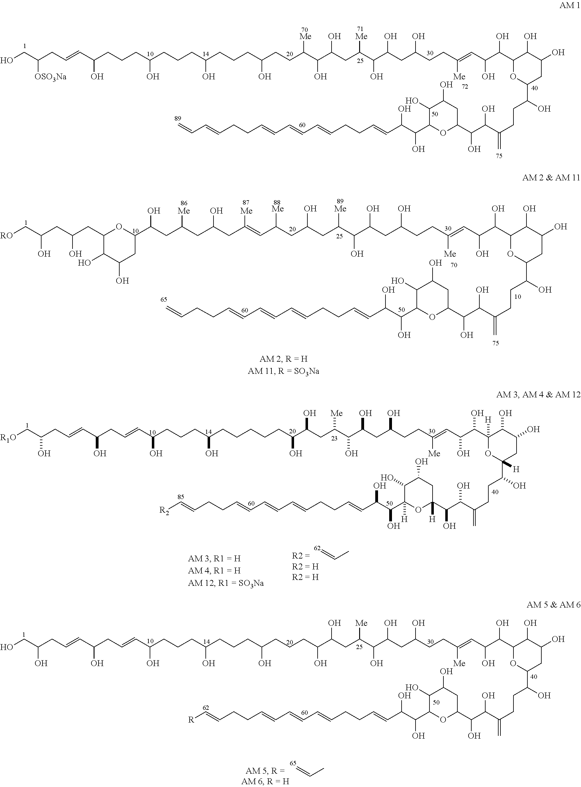
[0052]In order to evaluate the effects of the extract and amphidinol derivatives prepared in Example 1 on mycoplasma, Mycoplasma hyorhinis (ATCC17981s) was cultured in a PPLO medium (pleuropneumonia-like organism basal medium, Difco Laboratories, Detroit, USA) supplemented with a fetal calf serum and a yeast extract, according to the method disclosed by Hayflick (Hayflick, 1965, Tex. Rep. Bio. Med. 23, 285-303). That is, 100 μl of the medium was placed on each well of a 96-well plate, and a sample (that is, the extract and amphidinol derivatives prepared in Example 1) was added to each well, such that each sample was diluted from 2 times (i.e., 2× diluted) to 12 times (i.e., 12× diluted), the starting concentration being 1 mg / ml. Mycoplasma was cultured in a PPLO medium (pleuropneumonia-like organism basal medium, Difco Laboratories, Detroit, USA) supplemented with a fetal calf serum and a yeast extract, at a temperature o...
example 3
Removal of Mycoplasma from Cell Lines Contaminated with Mycoplasma
[0054](1) Collecting Cell Lines Contaminated with Mycoplasma
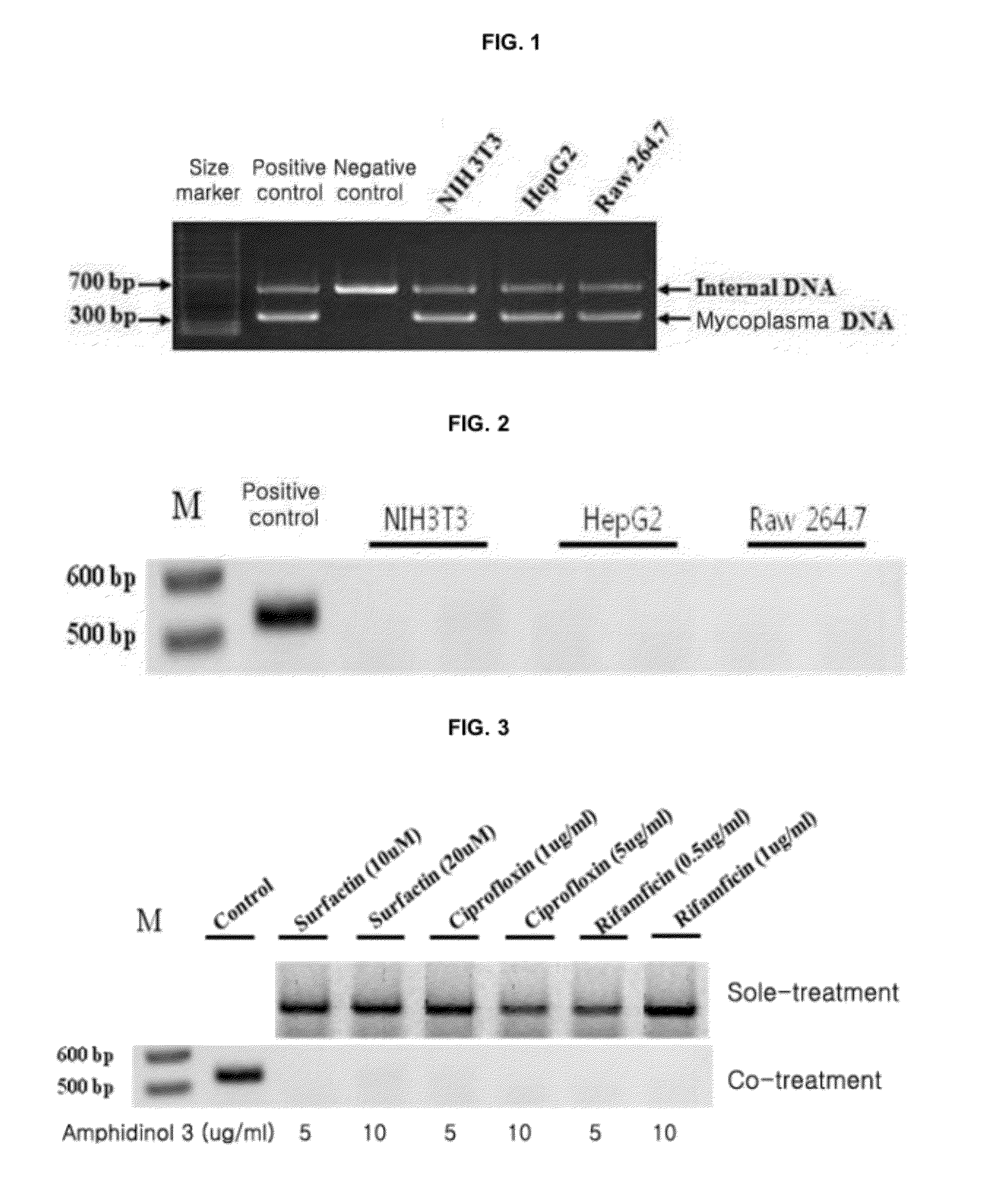
[0055]Various cell lines constructed in laboratories of colleges located in Seoul were obtained. The obtained cell lines were cultured in a DMEM (GIBCO BRL, Cat. No. 12800-058) for 3 days, and then the cells were harvested. PCR was performed thereon with a mycoplasma diagnosis kit (Promokine, Germany; Cellsafe Co., Ltd., Korea) according to the enclosed protocol. The resulting PCR products were electrophoresed on a 1% agarose gel to measure the mycoplasma contamination. As a result, it was confirmed that three cell strains, i.e., NIH3T3, HepG2, Raw 264.7 cell lines, were infected with mycoplasma (see FIG. 1).
[0056]The cell lines NIH3T3, HepG2, and Raw 264.7, which were confirmed to be contaminated with mycoplasma, were each inoculated at a concentration of 1×10⁶ cells on a T-75 culture flask (NUNC, Cat. No. 156367). DMEM (GIBCO BRL, Cat. No. 12800-058) contai...
example 4
Evaluation of Preventive Activity Against Mycoplasma Contamination During Cell Culturing
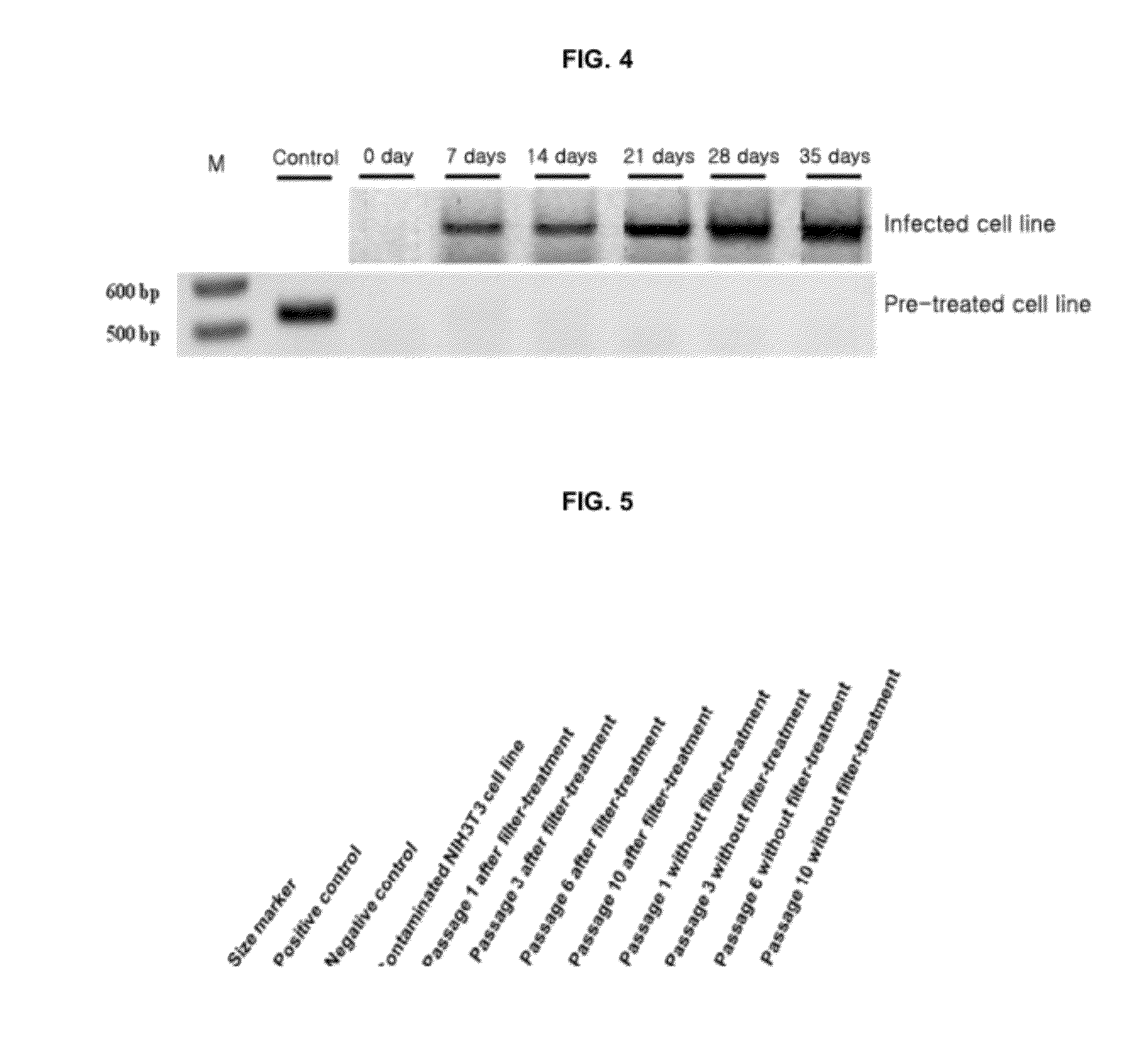
[0061]A cell line HepG2, which was not contaminated with mycoplasma, was inoculated at a concentration of 1×10⁵ cells on DMEM (GIBCO BRL, Cat. No. 12800-058) including 10% or less serum (GIBCO BRL, Cat. No. 10082-147). The cell line was treated with 20 μg / ml of amphidinol 3 (AM3) and 20 μM of surfactin, followed by inoculation of 1×10³ cfu (colony formin unit) of Mycoplasma hyorhinis (ATCC 17981). The cell line was cultured in an incubator at a temperature of 37° C. and in a condition of 5% CO2 for 3 days. Sub-culturing was performed twice per week. The cells were harvested every week and then PCR was performed thereon with a mycoplasma diagnosis kit (Promokine, Germany; Cellsafe Co., Ltd., Korea) according to the enclosed protocol. The resulting PCR products were electrophoresed on a 1% agarose gel to measure the mycoplasma contamination. Results thereof are as shown in FIG. 4. From the results ...
PUM
 Abstract
Description
Claims
Application Information
 - R&D
- Intellectual Property
- Life Sciences
- Materials
- Tech Scout
- Unparalleled Data Quality
- Higher Quality Content
- 60% Fewer Hallucinations
Browse by: Latest US Patents, China's latest patents, Technical Efficacy Thesaurus, Application Domain, Technology Topic, Popular Technical Reports.
© 2025 PatSnap. All rights reserved.Legal|Privacy policy|Modern Slavery Act Transparency Statement|Sitemap|About US| Contact US: help@patsnap.com


