Radiological image radiographing apparatus and method

a radiographing apparatus and radiographing technology, applied in the field of radiographing apparatus and method, can solve the problems of deteriorating image quality and down processing efficiency, and achieve the effect of preventing the deterioration of image quality of an image displayed based on the plurality of radiographs and simple operation

Inactive Publication Date: 2012-12-20
FUJIFILM CORP
View PDF4 Cites 2 Cited by
  • Summary
  • Abstract
  • Description
  • Claims
  • Application Information

AI Technical Summary

Benefits of technology

[0008]Since the above method disclosed in JP2001-092948A is a method of performing image processing on a radiological image, deterioration of the image quality of the generated image can be prevented. However, when the method disclosed in JP2001-092948A is applied to the generation of a tomographic image or a stereoscopic image based on a plurality of radiological images, processing efficiency is down. In addition, the method disclosed in JP2010-155017A only compares images using the index values of radiological images. In addition, the method disclosed in JP2008-026334A only compares images using information from histograms and the like in order to detect the position of a circuit pattern.
[0009]The present invention has been made in view of the above-mentioned problems and an object of the present invention is to prevent deterioration of the image quality of a displayed image when displaying one image such as a tomographic image or a stereoscopic image based on a plurality of radiological images acquired by performing radiographing multiple times in order to display a tomographic image or a stereoscopic image.
[0021]According to the aspect of the present invention, a plurality of radiological images are acquired, feature amounts indicating respective image qualities of the plurality of radiological images are calculated, the feature amounts of the plurality of radiological images are compared, and based on the comparison result, it is determined whether or not an abnormal radiological image with different image quality is present in the plurality of radiological images. For this reason, in a case where there is an abnormal radiological image, when a tomographic image is generated using a plurality of radiological images or a stereoscopic image is displayed, it is possible to do so without using the abnormal radiological image or to stop the display of a tomographic image or a stereoscopic image. As a result, deterioration of the image quality of an image displayed based on the plurality of radiological images can be prevented.
[0022]In addition, since the feature amounts are calculated based on any of the density histograms, spatial frequencies, edge information, S / N ratios, and contrast of the plurality of radiological images, the feature amounts can be calculated with a simple operation.
[0023]In addition, since a radiological image with feature amounts, which are different from those of many other radiological images by a predetermined threshold value or more, is identified as an abnormal radiological image, the presence of an abnormal radiological in the plurality of radiological images can be easily determined.

Problems solved by technology

Accordingly, if a tomographic image or a stereoscopic image is generated using such a radiological image, the image quality of a generated image deteriorates.
However, when the method disclosed in JP2001-092948A is applied to the generation of a tomographic image or a stereoscopic image based on a plurality of radiological images, processing efficiency is down.

Method used


Image

  • Radiological image radiographing apparatus and method
  • Radiological image radiographing apparatus and method
  • Radiological image radiographing apparatus and method

Examples


first embodiment

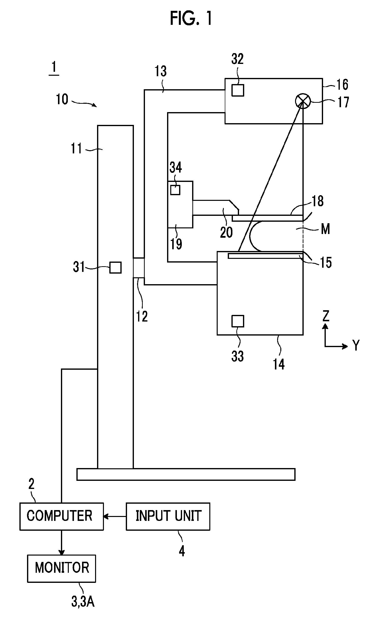
[0035]Hereinafter, embodiments of the present invention will be described with reference to the accompanying drawings. FIG. 1 is a view showing the schematic configuration of a radiological image radiographing apparatus according to the present invention. A radiological image radiographing apparatus 1 acquires a plurality of radiological images by radiographing a breast M from different radiographing directions in order to generate a tomographic image by performing tomosynthesis of the breast M and also to generate a stereo image for stereoscopic viewing of a radiological image of the breast M. As shown in FIG. 1, the radiological image radiographing apparatus 1 includes a radiographing unit 10, a computer 2 connected to the radiographing unit 10, and a monitor 3 and an input unit 4 connected to the computer 2.

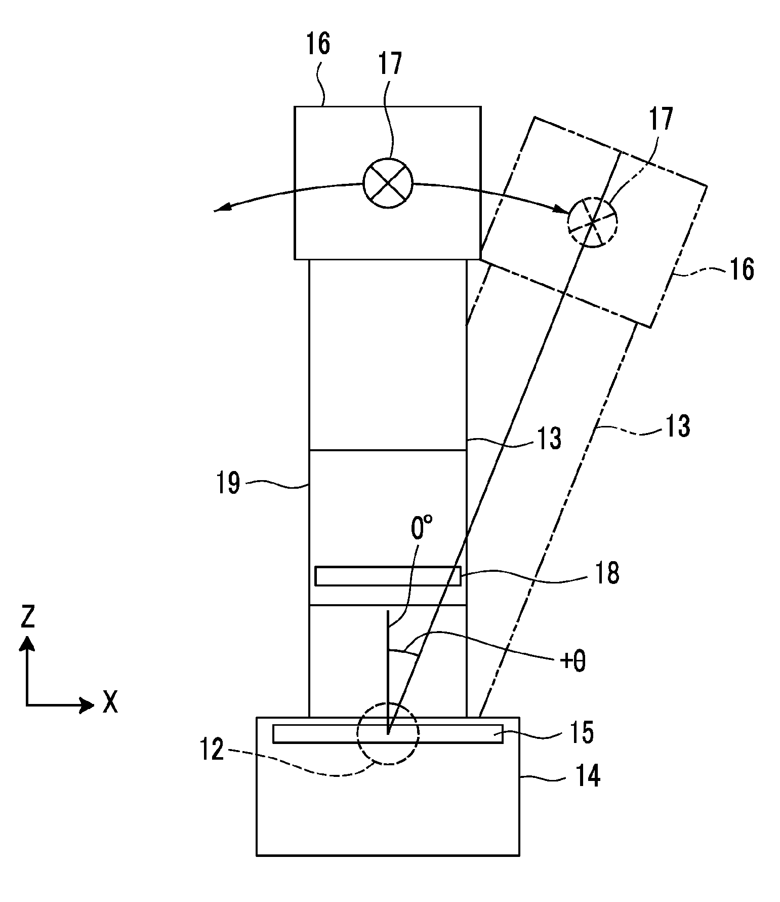
[0036]The radiographing unit 10 includes a pedestal 11, a rotary shaft 12 which can be rotated and moved up and down (in a Z direction) with respect to the pedestal 11, and an...

second embodiment

[0079]Thus, in the second embodiment, a radiological image with different image quality from others is not used for reconstruction since reconstruction is stopped when an abnormal radiological image is included in a plurality of radiological images. As a result, deterioration of the image quality of a tomographic image, which is caused by the generation of a tomographic image using an abnormal radiological image, can be prevented.

[0080]Next, a third embodiment of the present invention will be described. In addition, a radiological image radiographing apparatus according to a third embodiment has the same configuration as the radiological image radiographing apparatus according to the first embodiment except that a monitor 3A (display unit that displays a stereoscopic image) capable of performing three-dimensional display of a stereo image. Accordingly, detailed explanation thereof will be omitted herein. The radiological image radiographing apparatus according to the third embodimen...

third embodiment

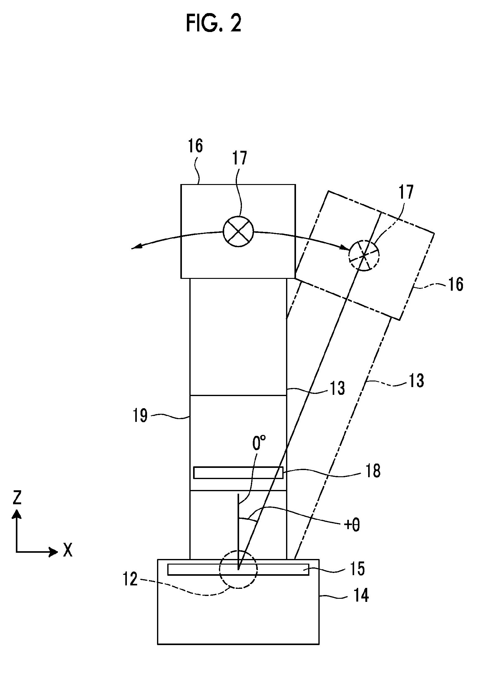
[0083]If there is an instruction to start radiographing through the input unit 4, radiographing (stereo radiographing) of two radiological images for displaying a stereo image is performed (step ST42). Specifically, first, the control unit 2a reads an angle of convergence θ1 for radiographing of a stereo image set in advance and outputs the information of the read angle θ1 to the arm controller 31. In addition, in the third embodiment, θ1=±4° is stored in advance as the information of the angle θ1 at this time. However, an operator may set an arbitrary angle of convergence through the input unit 4 without being limited to this.

[0084]Then, the arm controller 31 receives the information of the angle of convergence θ1 output from the control unit 2a. According to this information, first, the arm controller 31 outputs a control signal to rotate the position of the arm unit 13 by +θ1° with respect to the radiography platform 14. That is, in the third embodiment, the arm controller 31 out...


PUM

PropertyMeasurementUnit
densityaaaaaaaaaa
spatial frequenciesaaaaaaaaaa
thresholdaaaaaaaaaa

Abstract

Deterioration of the image quality of a displayed image is prevented when displaying one image, such as a tomographic image or a stereoscopic image, based on a plurality of radiological images acquired by performing radiographing multiple times in order to display a tomographic image or a stereoscopic image. A feature amount calculation unit calculates feature amounts representing the image quality, such as density histograms, for a plurality of radiological images for generating a tomographic image acquired by tomosynthesis. A determination unit compares the feature amounts of the plurality of radiological images and based on the comparison result, and then determines whether or not an abnormal radiological image with different image quality is present in the plurality of radiological images. When an abnormal radiological image is present, a reconstruction unit generates a tomographic image by performing reconstruction after excluding the abnormal radiological image from the plurality of radiological images.

Description

BACKGROUND OF THE INVENTION[0001]1. Field of the Invention[0002]The present invention relates to a radiological image radiographing apparatus and method for acquiring a plurality of radiological images by radiographing a subject from a plurality of different radiographing directions.[0003]2. Description of the Related Art[0004]An apparatus which displays a stereoscopic image (a three-dimensional image or a stereo image) based on two radiological images including information regarding parallax between both the left and right eyes has been proposed as an apparatus which displays a medical image, such as a radiological image. Such an apparatus irradiates a subject from different directions, detects radiation transmitted through the subject using a radiation detector to acquire a plurality of radiological images with parallax with respect to one another, and displays a stereoscopic image based on these radiological images that is able to be viewed stereoscopically. By displaying a stere...

Claims


Application Information

Patent Timeline
Patent Type & Authority Applications(United States)
IPC IPC(8): A61B6/03A61B6/02G01N23/04
CPCA61B6/022A61B6/025A61B6/0414A61B6/4452A61B6/464A61B6/502A61B6/5205A61B6/586G06T7/0002G06T2207/10072G06T2207/10116G06T2207/30168
Inventor NAKAYAMA, HIROKI
Owner FUJIFILM CORP
Why Eureka
Try Eureka