Drug Delivery and Substance Transfer Facilitated by Nano-Enhanced Device Having Aligned Carbon Nanotubes Protruding from Device Surface

a carbon nanotube and nano-enhanced technology, which is applied in the direction of prosthesis, treatment, application, etc., can solve the problems of affecting the implanting process, and affecting the effect of the implanting process

Active Publication Date: 2013-06-20
CALIFORNIA INST OF TECH
View PDF7 Cites 6 Cited by
  • Summary
  • Abstract
  • Description
  • Claims
  • Application Information

AI Technical Summary

Benefits of technology

[0018]The present invention relates to a nano-enhanced device for substance transfer between the device and a tissue. In one aspect, the device comprises a substrate and an array of substantially aligned carbon nanotubes having two ends, the carbon nanotubes being anchored within the substrate with at least one end protruding from the substrate, whereby the protruding carbon nanotubes enhance the substance transfer capabilities of the device.
[0023]In another aspect of the device, delivery of the drug from the device to the tissue is enhanced by a method selected from the group consisting of direct or indirect heating of at least a portion of the carbon nanotube array, electrical current through at least a portion of the carbon nanotube array, laser or other optical stimulation of at least a portion of the carbon nanotube array, ultrasonic waves on at least a portion of the carbon nanotube array, mechanical vibration of at least a portion of the carbon nanotube array, and modifying the hydrophobicity of the nanotube array during fabrication.
[0025]In another aspect of the device of the present invention, the carbon nanotubes are arranged in one or more clusters, such that each cluster effectively functions as a needle.
[0030]In a further aspect of the present invention, a subset of the carbon nanotube clusters is treated with glucose oxidase, and another subset of the carbon nanotubes is treated with ferric cyanide, whereby conduction through the blood may be achieved and allows for measurement of the blood-glucose level.

Problems solved by technology

Use of such a needle is typically associated with pain, exposure of the blood to the environment, or risk of infection due to the rupture of the skin.
Again, this procedure (depending on the scale) to achieve the implant is problematic both due to the process of placing the implant and the potential rejection by the patient's body.
The use of an expanded balloon to forcibly open the narrowed section of the artery requires very high pressure (15 bars) and tends to cause injury to the vessel walls.
The body's natural response to such injury is hyperproliferation, an abnormally high rate of cell division, which results in lumen narrowing and thus decreased function of the vessel.
This is counterproductive to the initial goal of opening the stenotic or occluded artery.
The mechanism of action of paclitaxel is the stabilization of microtubules through binding to tubulin, thus interfering with their normal breakdown during cell division.
1. The need of long inflation time in order to make sure the paclitaxel diffusion to the arterial wall is sufficient, which may cause excessive arterial wall injury. Previous study shows that 40-60 minutes are required to transfer roughly 90% of the initial dose of paclitaxel to the arterial wall (See Literature Reference No. 1);
2. In order to obtain the optimum benefit of hyperproliferation inhibition, it is important that the paclitaxel coating is not lost or washed off by the blood stream while advancing into the stenotic segment of the artery. Previous study shows that the rate of paclitaxel being washed away from the uninflated balloon by the blood stream is up to 1.2% from the initial dose per minute (See Literature Reference No. 2);
3. Potential loss of paclitaxel coating due to contact with the healthy arterial wall while advancing into the stenotic segment of the artery; and
4. The maximum dose of paclitaxel that can be applied on a single balloon is around 11 mg (10 μg / mm2), which is significantly less than the paclitaxel dose approved by the FDA for Taxol (See Literature Reference No. 1).
However, in that patent, the carbon nanotubes were mixed with the base material for the purpose of strengthening the material and not for drug delivery purposes.
In addition, the carbon nanotubes were not aligned or partially anchored in the base material in a way such that they would protrude from the surface and facilitate drug delivery, rendering the device non-functional for that purpose.

Method used


Image

  • Drug Delivery and Substance Transfer Facilitated by Nano-Enhanced Device Having Aligned Carbon Nanotubes Protruding from Device Surface
  • Drug Delivery and Substance Transfer Facilitated by Nano-Enhanced Device Having Aligned Carbon Nanotubes Protruding from Device Surface
  • Drug Delivery and Substance Transfer Facilitated by Nano-Enhanced Device Having Aligned Carbon Nanotubes Protruding from Device Surface

Examples


Embodiment Construction

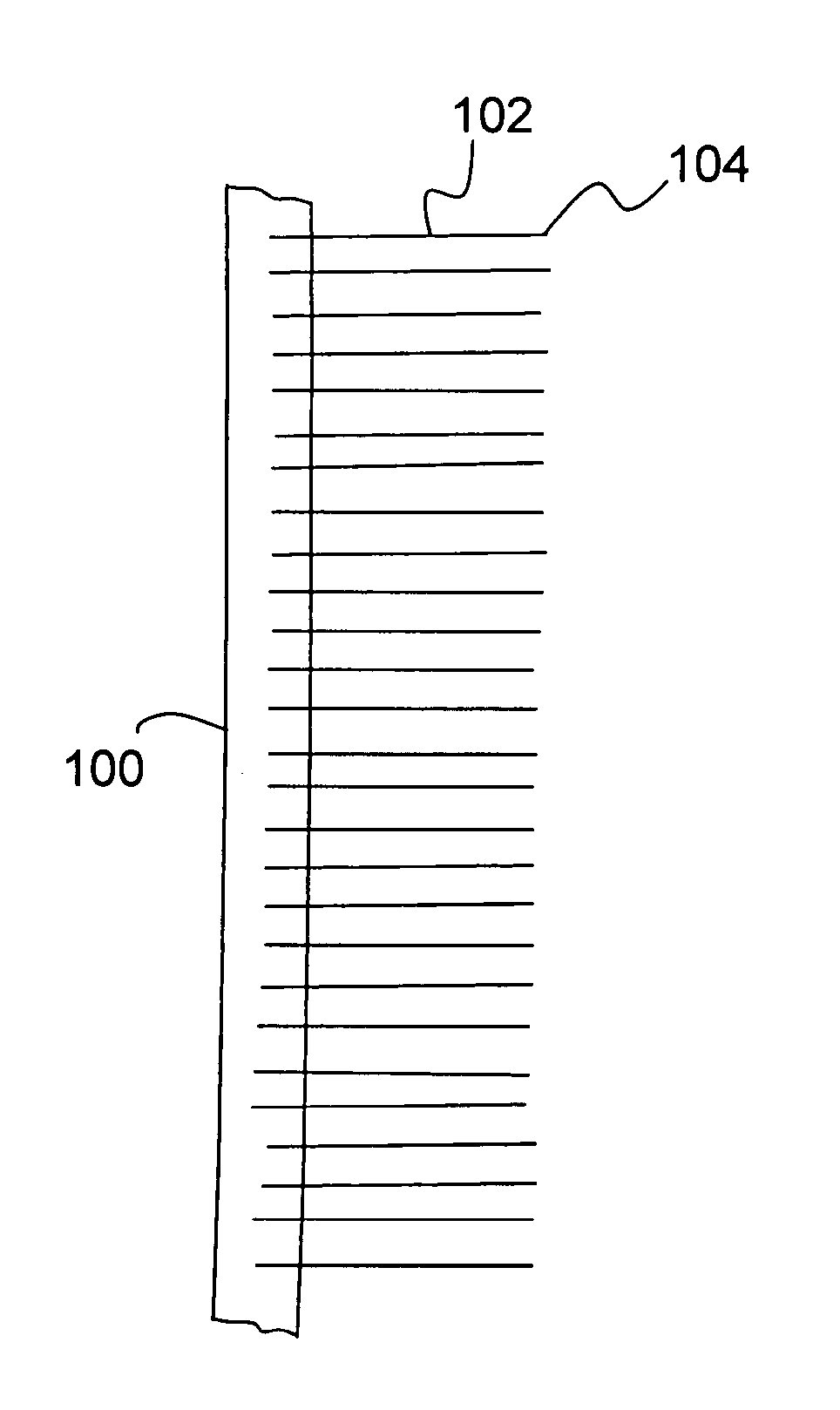
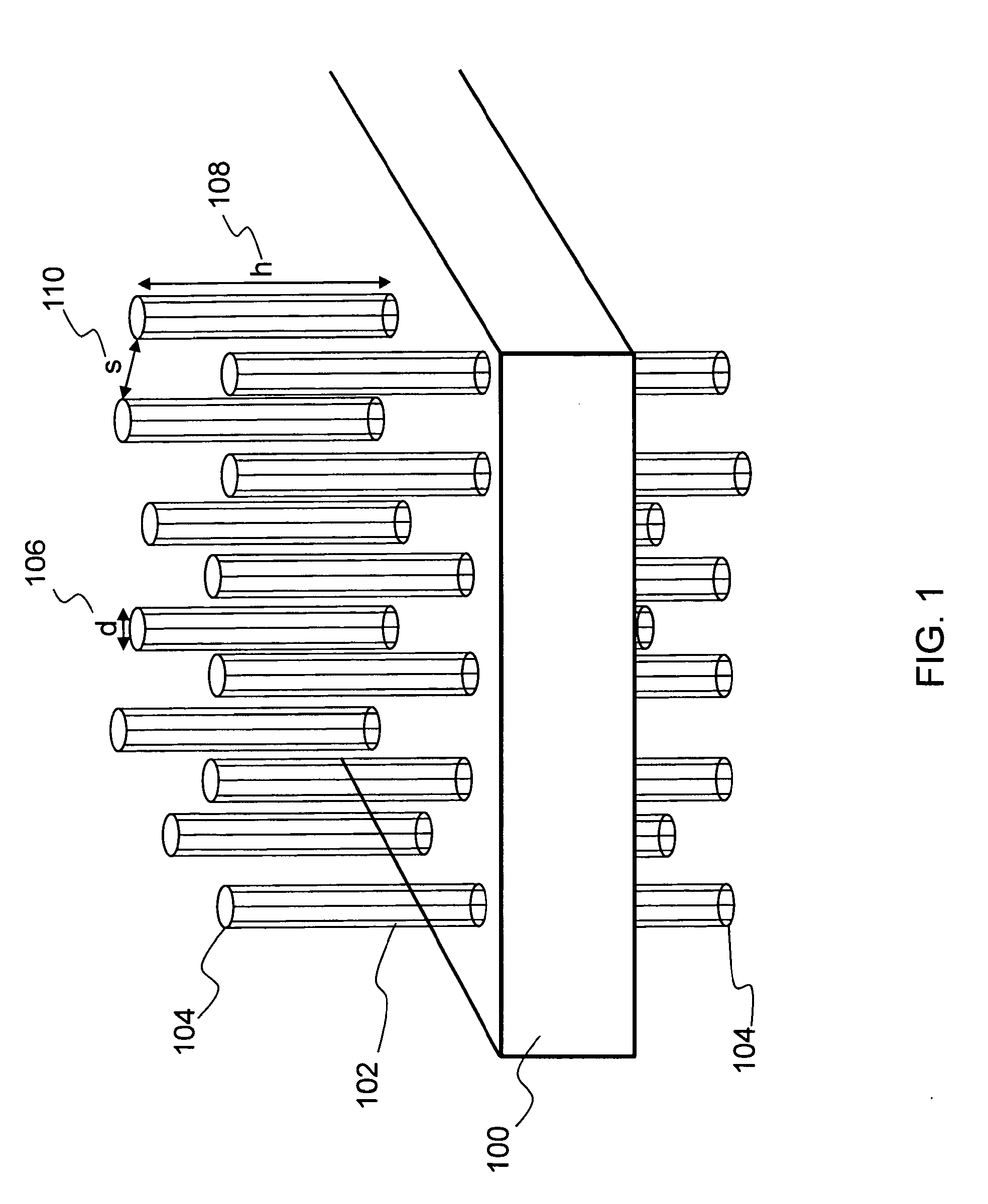
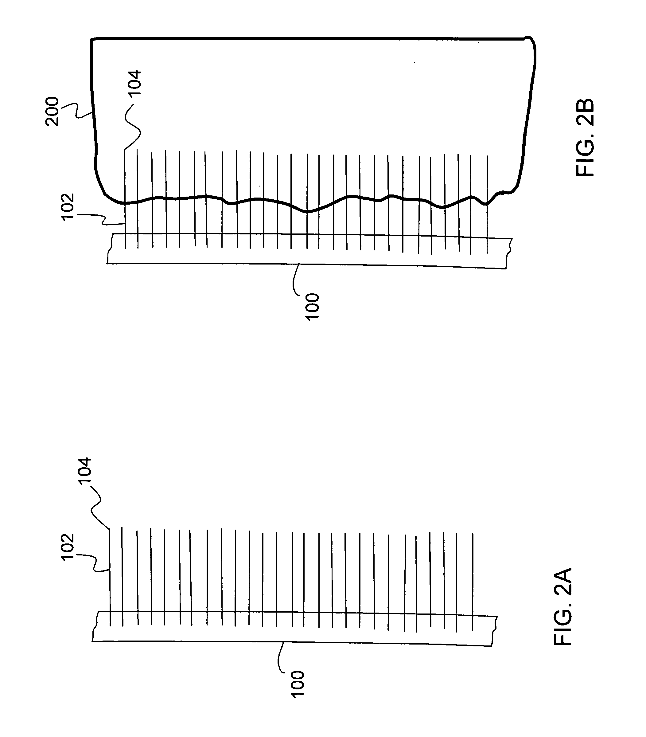
[0045]The present invention relates to a nanostructure-enhanced drug delivery and blood monitoring device and, more particularly, to a device with aligned carbon nanotubes protruding from the device surface for facilitating drug or fluid transfer between the device and body tissue. The following description is presented to enable one of ordinary skill in the art to make and use the invention and to incorporate it in the context of particular applications. Various modifications, as well as a variety of uses in different applications will be readily apparent to those skilled in the art, and the general principles defined herein may be applied to a wide range of embodiments. Thus, the present invention is not intended to be limited to the embodiments presented, but is to be accorded the widest scope consistent with the principles and novel features disclosed herein.

[0046]In the following detailed description, numerous specific details are set forth in order to provide a more thorough u...


PUM

PropertyMeasurementUnit
diameteraaaaaaaaaa
diameteraaaaaaaaaa
temperatureaaaaaaaaaa

Abstract

The present invention relates to a nano-enhanced device for substance transfer between the device and a tissue. The device comprises a substrate with substantially aligned carbon nanotubes anchored within the substrate, and with at least one end of the carbon nanotubes protruding from the substrate. The protruding nanotube ends may be coated with a drug for delivery of the drug into body tissue. The present invention may be incorporated into an angioplasty catheter balloon or into a patch that is worn on the skin. The carbon nanotubes can be grouped in clusters to effectively form nano-needles which can transfer fluid to or from the subdermal tissue. The nano-needles can be used in conjunction with a sensor to ascertain body fluid information such as pH, glucose level, etc.

Description

PRIORITY CLAIM[0001]This is a continuation of U.S. patent application Ser. No. 12 / 657,862, filed on Jan. 27, 2010, entitled “DRUG DELIVERY AND SUBSTANCE TRANSFER FACILITATED BY NANO-ENHANCED DEVICE HAVING ALIGNED CARBON NANOTUBES PROTRUDING FROM DEVICE SURFACE,” which is hereby incorporated by reference in its entirety. This application is also a continuation-in-part of U.S. patent application Ser. No. 11 / 827,169, filed on Jul. 10, 2007, entitled “METHOD FOR SELECTIVELY ANCHORING LARGE NUMBERS OF NANOSCALE STRUCTURES.” This application also is a non-provisional patent application claiming benefit of priority of U.S. Provisional Application No. 61 / 206,071, filed on Jan. 27, 2009, entitled “CARBON NANOTUBE-BASED DEVICE FOR SUB-DERMAL BLOOD AND FLUID PROPERTY MONITORING,” and U.S. Provisional Application No. 61 / 179,639, filed on May 19, 2009, entitled, “METHOD, DEVICE AND DESIGN FOR DELIVERY OF DRUGS BY A NANO-ENHANCED ANGIOPLASTY BALLOON.”FIELD OF INVENTION[0002]The present invention ...

Claims


Application Information

Patent Timeline
Patent Type & Authority Applications(United States)
IPC IPC(8): A61M37/00A61N5/06A61M5/44A61B5/145A61M25/10A61N1/30A61F2/958
CPCA61K47/48992A61M2037/0053A61L27/54A61L27/56A61L2300/258A61L2300/416A61L2300/602A61L2300/606A61L2400/12B29C70/64B29K2105/162B82Y5/00B82Y30/00B82Y40/00C01B31/0206A61B5/14532A61M5/44A61M25/104A61M37/0015A61M37/0092A61N1/306A61N5/062A61M2037/0023A61L27/443C01B32/15A61K47/6957A61M2037/0061
Inventor GHARIB, MORTEZAARIA, ADRIANUS I.SANSOM, ELIJAH BODHI
Owner CALIFORNIA INST OF TECH
Why Eureka
Try Eureka