Methods and compositions for preparing samples for immunostaining

a technology of immunostaining and composition, which is applied in the field of immunostaining preparation methods and compositions, can solve the problems of degrading the cellular morphology of cytology specimens, high heat pretreatment, etc., and achieves the effect of maintaining cellular morphology

Active Publication Date: 2013-06-27
TRIPATH IMAGING INC
View PDF17 Cites 2 Cited by
  • Summary
  • Abstract
  • Description
  • Claims
  • Application Information

AI Technical Summary

Benefits of technology

[0006]Compositions and methods for preparing samples for immunostaining are provided herein. Compositions disclosed herein include kits comprising a first solution comprising a surfactant and a second solution comprising a chaotropic agent. The two solutions find use in retrieving protein epitopes by sequentially contacting a sample, such as cells or tissues, with the first solution and then the second solution. The exposed antigen is then available for binding to an antibody, which can be detected using any method known in the art. The presently disclosed methods and compositions allow for antigen retrieval in the absence of extreme heat, thus maintaining cellular morphology.

Problems solved by technology

Cytology specimens are not fixed to the same degree as tissue samples, are not normally embedded in paraffin, and do not contain stromal support material to maintain cellular morphology during high heat pretreatment.
Thus, high heat pretreatment methods that serve to increase epitope exposure and accessibility to the primary antibody degrade the cellular morphology of cytology specimens.

Method used


Image

  • Methods and compositions for preparing samples for immunostaining
  • Methods and compositions for preparing samples for immunostaining
  • Methods and compositions for preparing samples for immunostaining

Examples


example 1

Analysis of Antigen Retrieval Solutions and Incubation Temperatures

[0177]Historically, most tissue-based antigen retrieval (AR) techniques have utilized high heat, ˜100° C. and / or treatment with various solutions. The AR procedure for the detection of the antigens MCM2 and MCM7 in SurePath® (TriPath Imaging, Inc.) cervical cytology samples using a cocktail of three antibodies disclosed in U.S. Pat. Nos. 7,595,380 (27C5.6 and 26H6.19 anti-MCM2 antibodies) and 7,632,498 (2E6.2 anti-MCM7 antibody), which are herein incorporated by reference in their entirety, was optimized as described hereinbelow to maintain cell morphology, while ensuring appropriate immunostaining of cytology specimens by investigating multiple AR solutions and incubation temperatures.

[0178]A broad range of potential AR (pretreatment) solutions (n=43) were tested on SurePath® cytology specimens to determine which ones would have a positive effect on immunostaining with a triple antibody cocktail (two anti-MCM2 antib...

example 2

Comparison of the Effects of the Two-Step Pretreatment Method with a High-Heat Pretreatment Method on Immunostaining and Morphology in Cervical Cytology Samples

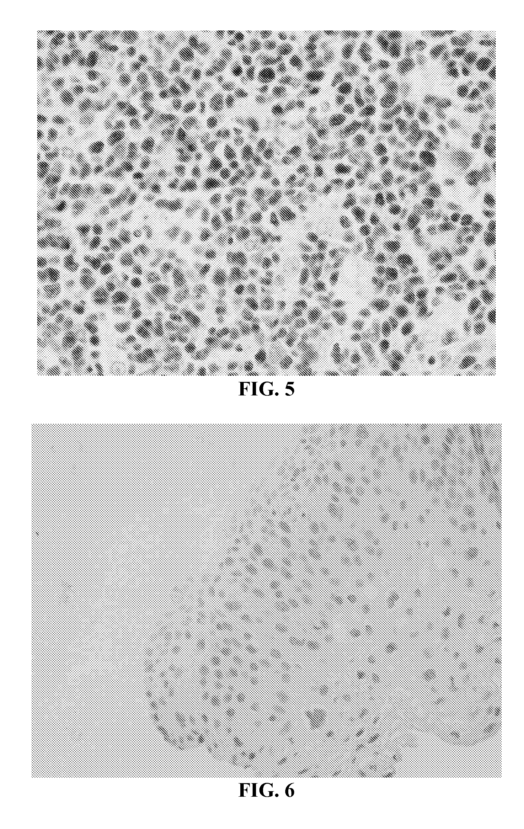
[0192]This study compared the two-step pretreatment method using two different chaotropic salt solutions for the second incubation step to each other and to a pretreatment method utilizing high heat (i.e., the steamer method described in Example 1).

[0193]In these experiments, SurePath® cervical cytology specimens were processed using the internally developed PrepStain Plus® Instrument (Tripath Imaging, Inc.) and deposited for staining. For those samples treated with the two-step pretreatment method, the slides were heated to 50° C. on specially-designed slide heating trays and Pretreatment Solution 1 (0.1% SDS) was applied and the slides were incubated at 50° C. for 19 minutes. The cells were washed with a standard Tris-buffered saline (TBS) solution. Pretreatment Solution 2 (either 3M LiClO4 / 0.1% NP-40 or 3M guanidine thiocy...

example 3

Detection of Antigens Ki67 and p16 in Cervical Cytology Samples Using the Two-Step Pretreatment Process

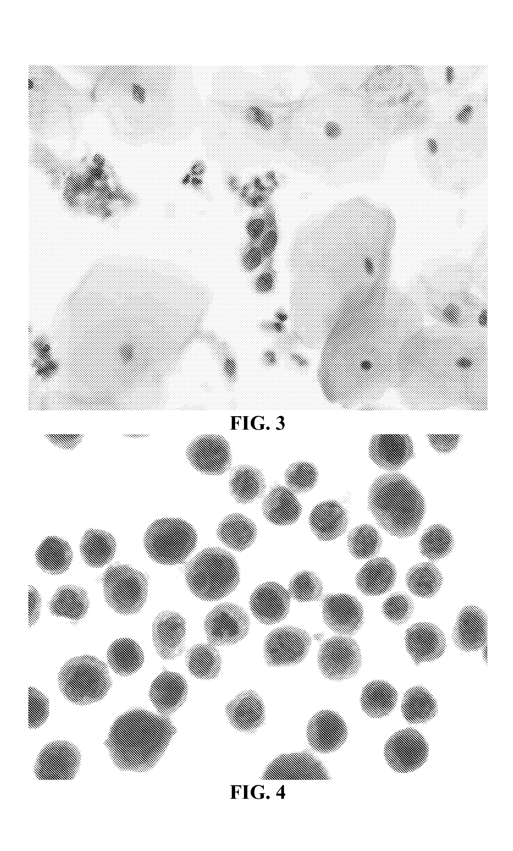
[0201]In these experiments, SurePath® cervical cytology specimens were processed using the internally developed PrepStain Plus® Instrument (Tripath Imaging, Inc.) and deposited for staining. Following cell deposition, the slides were heated to 50° C. on specially-designed slide heating trays and AR Solution 1 (0.1% SDS) was applied and the slides were incubated at 50° C. for 19 minutes. The cells were washed with a standard tris-buffered saline (TBS) solution. AR Solution 2 (3M LiClO4 / 0.1% NP-40) was then added and the cells were incubated an additional 19 minutes at 50° C. Following this incubation, the slides were washed again with TBS and processed for immunostaining and PAP counterstaining on the PrepStain Plus® instrument. The slides were then coverslipped and examined by a cytologist and / or cytopathologist. Representative results of cervical cytology samples immunostained wit...


PUM

PropertyMeasurementUnit
temperaturesaaaaaaaaaa
pHaaaaaaaaaa
temperatureaaaaaaaaaa

Abstract

Compositions and methods for preparing a sample for immunological staining are provided. Compositions include kits comprising a first solution comprising a surfactant and a second solution comprising a chaotropic agent. Methods comprise contacting a sample, such as cells or tissues, with a first solution comprising a surfactant and then contacting the sample with a second solution comprising a chaotropic agent. The method does not require extreme heat for antigen retrieval and therefore, maintains the cellular morphology of the sample.

Description

CROSS-REFERENCE TO RELATED APPLICATIONS[0001]This application claims the benefit of U.S. Provisional Application No. 61 / 555,139, filed Nov. 3, 2011, which is herein incorporated by reference in its entirety.FIELD OF THE INVENTION[0002]The present invention relates to methods and compositions to enhance the detection of proteins by making epitopes more accessible for antibody binding.BACKGROUND OF THE INVENTION[0003]Fixation of tissue and cellular samples often makes protein-based epitopes inactive, or inaccessible for immunostaining due to protein cross-linking. Antigen retrieval (AR) is the process by which target epitopes are made accessible for immunostaining. Overcoming fixation-induced cross-linking allows for target epitopes buried within the tertiary structure of proteins to become accessible for binding with primary antibodies. Antigen retrieval also advantageously lowers the threshold for detection of antigens, thereby reducing the amount of antibody needed for detection, r...

Claims


Application Information

Patent Timeline
Patent Type & Authority Applications(United States)
IPC IPC(8): G01N1/30
CPCG01N1/30G01N33/56966G01N33/57492
Inventor FISCHER, TIMOTHY J.NELSON, RAMONA R.TAYLOR, ADRIANN J.WHITEHEAD, CLARK M.
Owner TRIPATH IMAGING INC
Why Eureka
Try Eureka