Rapid microbial detection and antimicrobial susceptibility testing

a technology of microbial susceptibility testing and microbial detection, which is applied in the direction of biochemistry apparatus and processes, instruments, material analysis, etc., can solve the problems of not being able to provide the attending physician with specific diagnostic information, unable to significantly improve the outcomes of approximately 87% of patients so treated, and unable to change from ineffective to effective therapy.

Active Publication Date: 2013-08-22
ACCELERATED MEDICAL DIAGNOSTICS INC
View PDF29 Cites 56 Cited by
  • Summary
  • Abstract
  • Description
  • Claims
  • Application Information

AI Technical Summary

Problems solved by technology

Thus the standardized current procedures require at least two sequential growth cycles, each typically requiring many hours to complete.
It is well recognized in the medical community that in certain circumstances such methods, which attempt to prove the absence of growth, produce results that do not correlate adequately with the actual results of treatment.
As a result of these and other serious deficiencies, contemporary practice fails to provide the attending physician with the specific diagnostic information that the physician needs in order to select an effective drug to treat the infection within the desired time window.
That is, unless the physician initiates effective treatment, i.e. antimicrobial drugs of a type and concentration adequate to quickly kill the infectious organisms within substantially less than 24 hours from symptom onset, a change from ineffective to effective therapy will not significantly improve outcomes for approximately 87% of patients so treated.
Clinical research has demonstrated that such empiric drug treatment is ineffective in approximately 25% to 50% of cases.
Additionally, exposure of a patient to inadequate therapy not only increases the individual patient's costs and medical risks, but also increases the likelihood of fostering the emergence of resistant organisms.
The latter problem increases the medical risk not only for the individual patient, but for all other individuals in the hospital and community who may later become infected with resistant organisms.
It is well recognized in the clinical research literature that prior exposure of a patient to ineffective antibiotics constitutes a significant risk factor in the later emergence of resistant organisms in that patient.
However, none yet exist for routine bacterial identification, quantitation, and drug susceptibility testing for many serious infectious diseases.
In certain circumstances, some of these provide results in approximately 24 hours for a limited set of particular organisms.
However all commercial products still require sample enrichment by means of bacterial culturing before applying the tests.
However, the sensitivity of devices so far described in the literature for food testing or bio-defense falls far short of the requirements for medical diagnostics.
Furthermore, these non-diagnostic applications do not require drug susceptibility testing and so the aforesaid devices do not provide it nor apparently do they lend themselves to adaptation for such a purpose.
A key limitation with these devices and with laboratory methods such as ELISA is their dependency on the target analyte concentration.
This problem is exacerbated by the very low diffusion rates of bacteria.
These systems, however, suffer from a number of limitations, including the need to sequentially immobilize probes on the addressable electrodes, the need to perform sequential reactions, and limitations on the detection methods that can be employed due to the higher voltages that are required for electrophoresis, precluding the use of transparent electrodes (e.g. through the use of indium tin oxide), that cannot operate at the voltages used by the Nanogen system.
Furthermore, the higher voltages at which the Nanogen system operate generate oxidation products that are potentially harmful to the probes or targets, and which therefore requires the use of complex passivation surfaces to protect the probes and targets.
It is difficult with these systems to determine susceptibility to antimicrobial agents, given the difficulty of finding nucleic acid or antibody markers that reliably correlate with antimicrobial resistance or behavior.

Method used


Image

  • Rapid microbial detection and antimicrobial susceptibility testing
  • Rapid microbial detection and antimicrobial susceptibility testing
  • Rapid microbial detection and antimicrobial susceptibility testing

Examples


example 1

Electrokinetic Concentration

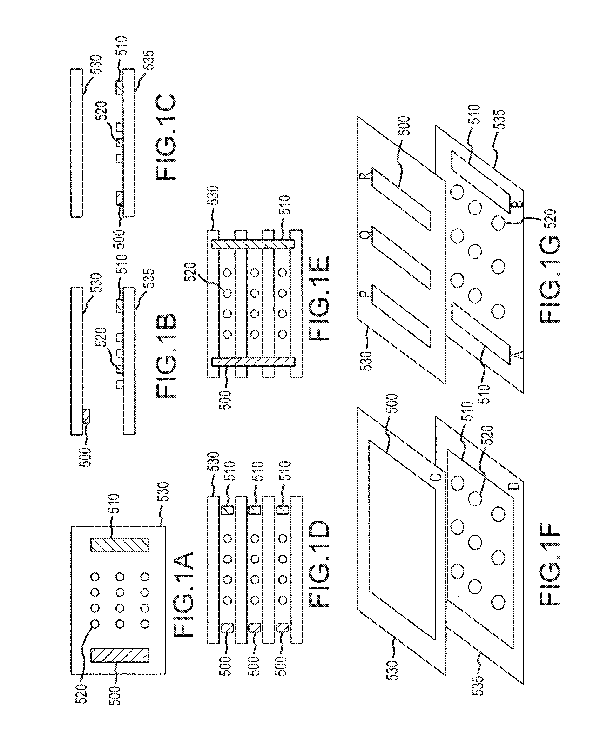
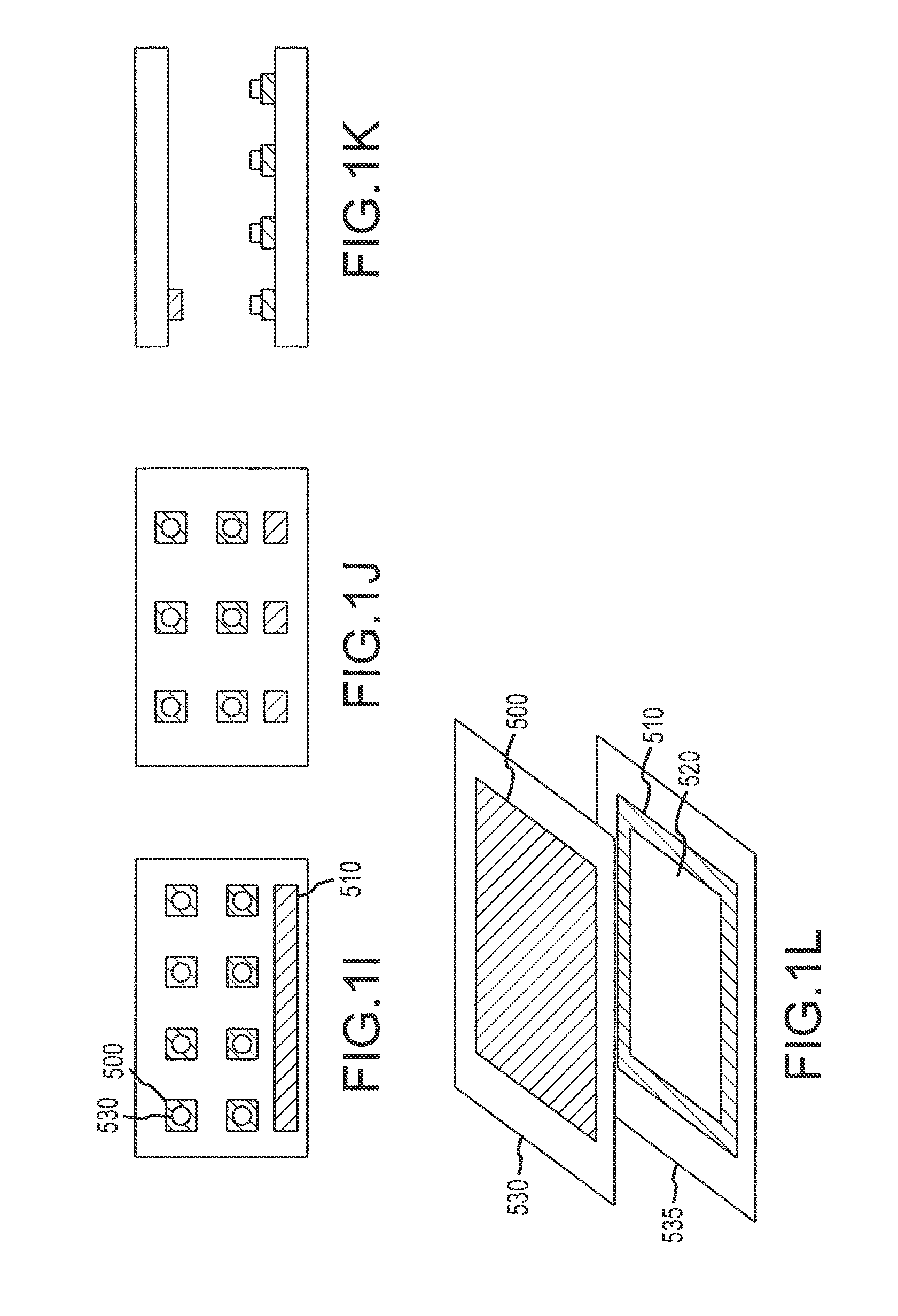
[0365]A flow cell was constructed by sandwiching two ITO modified glass microscope slides between a plastic laminate structure forming a 4 channel flow cell as illustrated in FIG. 50. The distance between the slide surfaces is approximately 500 microns. The bottom ITO slide was precoated with amine reactive OptiChem (Accelr8) and chemically modified with diethylene triamine (DETA) to yield a cationic bacteria capture surface.

[0366]A mixture of 1×10⁷ CFU / ml of Staphylococcus aureus in 10 mM hydroquinone, 10 mM DTT, and 10 mM CAPS pH 6.5 was injected into a flow chamber. A potential difference (1.6 V) was applied across the ITO slides for approximately 3 minutes to surface concentrate and bind the bacteria to the DETA OptiChem modified ITO electrode. Photomicrographs of the surface of the anode before the application of the potential and 50 seconds later show the presence of the electrokinetically concentrated S. aureus microorganisms. The number of bacteri...

example 2

Microorganism Growth

[0367]The redox solution from Example 1 was replaced with tryptic soy growth media and incubated at 37 C on an inverted microscope to establish detectable growth of bacteria bound to DETA surface. Photomicrographs of the S. aureus at the beginning of growth and after different incubation times were taken (data not shown). The photomicrographs were taken with phase optics, and the flare surrounding the bacteria indicated the presence of additional bacteria resulting from cell division of the original bacteria. Thus bacterial growth ca follow electrokinetic concentration.

example 3

AOA Susceptibility

[0368]Bacteria concentrated and grown as in Examples 1 and 2 were challenged by 5 micrograms / ml of oxacillin to which the S. aureus strain used was susceptible. Simultaneous with the administration of oxacillin was the administration of 10 micromolar propidium iodide, which is excluded from live cells, and thus whose presence in fluorescence images is indicative of cell death. Photomicrographs of the bacteria at the beginning of AOA treatment were taken, under phase contrast and fluorescence microscopy, and additional images were taken one hour later (data not shown). There was a large increase in fluorescence which indicated the large amount of cell death from the administration of oxacillin.


PUM

PropertyMeasurementUnit
timeaaaaaaaaaa
volumeaaaaaaaaaa
volumeaaaaaaaaaa

Abstract

A method for the detection of microorganisms in a sample comprising contacting said sample with a biosensor concentration module, allowing microorganisms to grow for a first period of time and detecting growth of discrete microorganisms as an indication of the presence of said microorganisms.

Description

CROSS-REFERENCE TO RELATED APPLICATIONS[0001]This application is a Continuation of U.S. application Ser. No. 13 / 311,100, filed Dec. 5, 2011, which application is a Continuation of U.S. application Ser. No. 12 / 021,087, filed Jan. 28, 2008, now U.S. Pat. No. 8,071,319, issued Dec. 6, 2011; which application is a Continuation of U.S. application Ser. No. 11 / 303,803, filed Dec. 16, 2005, now U.S. Pat. No. 7,341,841, issued Mar. 11, 2008; which application claims the benefit of U.S. Application Ser. No. 60 / 637,423, filed Dec. 16, 2004 and U.S. Application Ser. No. 60 / 638,989, filed Dec. 22, 2004; and[0002]U.S. application Ser. No. 11 / 303,803, filed Dec. 16, 2005, now U.S. Pat. No. 7,341,841, issued Mar. 11, 2008, is a Continuation-In-Part of U.S. application Ser. No. 10 / 888,828, filed Jul. 8, 2004, now U.S. Pat. No. 7,687,239, issued Mar. 30, 2010; which application claims the benefit of U.S. Application Ser. No. 60 / 486,605, filed Jul. 12, 2003 and U.S. Application Ser. No. 60 / 571,479, f...

Claims


Application Information

Patent Timeline
Patent Type & Authority Applications(United States)
IPC IPC(8): C12Q1/18G06F19/10G16B99/00
CPCC12Q1/04G06F19/10C12Q1/18G16B99/00
Inventor METZGER, STEVEN W.HOWSON, DAVID C.GOLDBERG, DAVID A.BUTTRY, DANIEL A.
Owner ACCELERATED MEDICAL DIAGNOSTICS INC
Why Eureka
Try Eureka