Methods for Rapid Distinction between Debris and Growing Cells

a technology of growing cells and debris, applied in biochemistry apparatus and processes, laboratory glassware, instruments, etc., can solve the problems of difficult miniaturization, high cost, and bulky optics of conventional optical microscopes, and neither can work well with confluent cell cultures or any sampl

Active Publication Date: 2014-05-15
CALIFORNIA INST OF TECH
View PDF56 Cites 99 Cited by
  • Summary
  • Abstract
  • Description
  • Claims
  • Application Information

AI Technical Summary

Problems solved by technology

Conventional optical microscopes have bulky optics, and have proven to be expensive and difficult to miniaturize.
However, neither can work well with confluent cell cultures or any sample in which cells are contiguously connected over a sizable length scale.
Adherent, confluent, or contiguously arranged specimens are usually incompatible with imaging in a fluidic mode.
In addition, the field of view may be limited by the geometry of the fluid channel.
However, such approaches appear to have not been applied to targets that occupy more than 0.1 mm2 in total contiguous area coverage with submicron resolution, as found in Repetto, Madanyali, Xu, Garcia-Sucerquia, and also in Biener, G., et al., “Combined reflection and transmission microscope for telemedicine applications in field settings,” Lab Chip, Vol. 11, pp.
The reason for this limitation is well-known: the loss of phase information during the intensity recording process.
The generated images based on these mitigating methods have artifacts that may arise from interference, and are identifiably different and of lower quality than images acquired with conventional microscopes due to coherence based noise sources.

Method used


Image

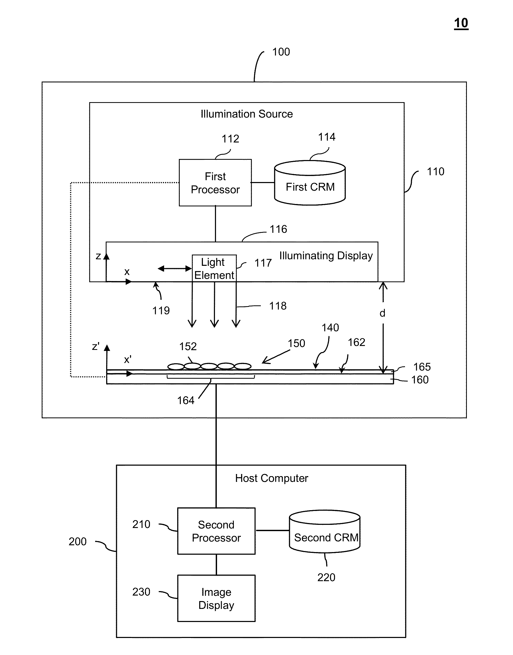
  • Methods for Rapid Distinction between Debris and Growing Cells
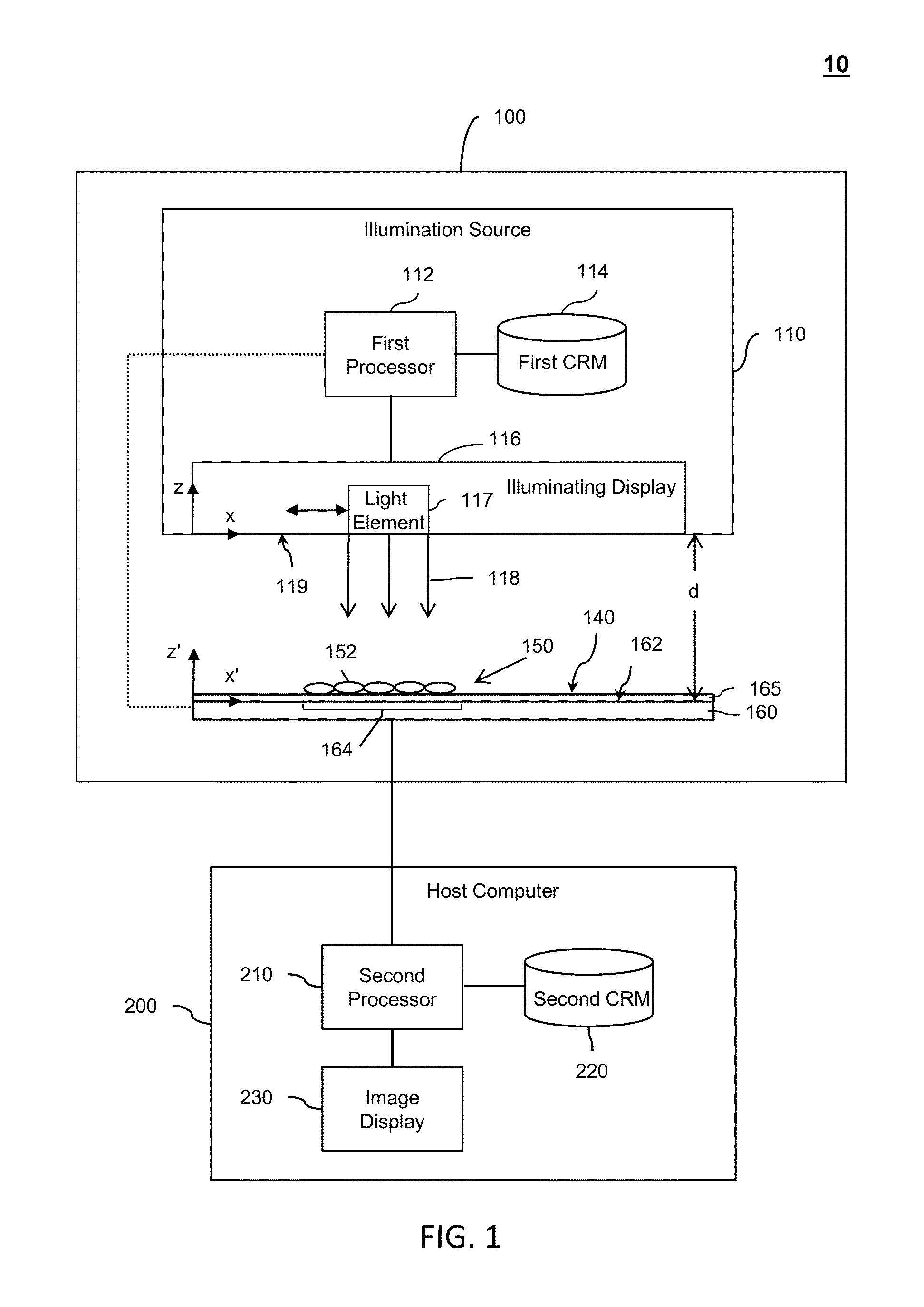
  • Methods for Rapid Distinction between Debris and Growing Cells
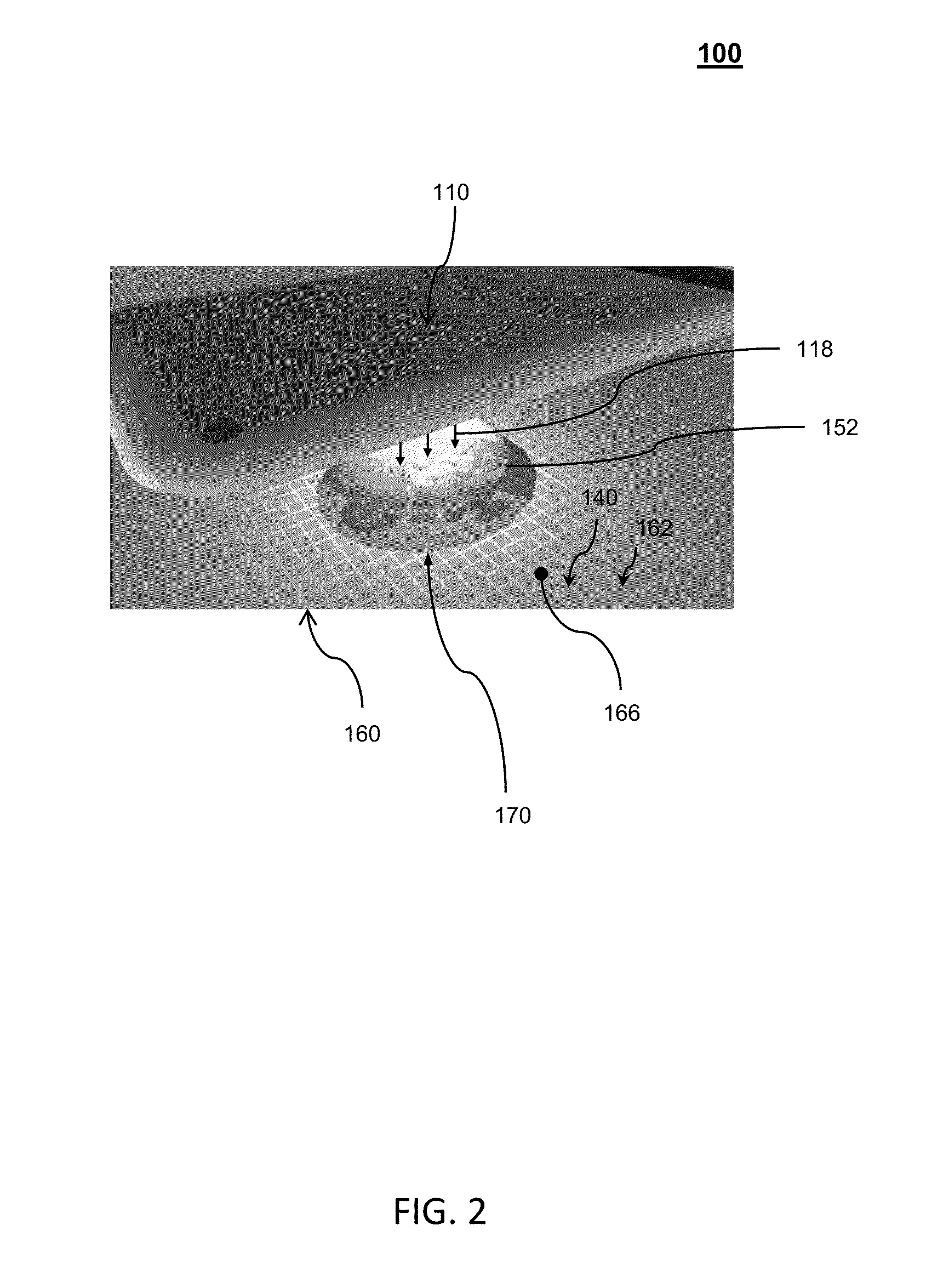
  • Methods for Rapid Distinction between Debris and Growing Cells

Examples


Embodiment Construction

[0048]The need for a high-resolution, wide-field-of-view, cost-effective microscopy solution for autonomously imaging growing and confluent specimens (e.g., cell cultures), especially in longitudinal studies, is a strong one, as discussed in Schroeder, T., “Long-term single-cell imaging of mammalian stem cells,” Nature Methods 8, pp. S30-S35 (2011), which is hereby incorporated by reference in its entirety for all purposes. Some examples of experiments that would benefit from such a solution include: 1) the determination of daughter fates before the division of neural progenitor cells, as discussed in Cohen, A. R., Gomes, F. L. A. F., Roysam, B. & Cayouette, M., “Computational prediction of neural progenitor cell fates,” (2010); 2) the existence of haemogenic endothelium, as discussed in Eilken, H. M., Nishikawa, S. I. & Schroeder, T., “Continuous single-cell imaging of blood generation from haemogenic endothelium,” Nature 457, pp. 896-900 (2009), 3) neural and hematopoietic stem an...


PUM


Abstract

Methods of rapid distinction between growing cells and debris, which determine a time-lapse movie of specimen images, track features of each entity, and categorize each entity as growing cells or debris.

Description

CROSS-REFERENCES TO RELATED APPLICATIONS[0001]The present application is a continuation-in-part application of U.S. patent application Ser. No. 13 / 411,302 titled “E-PETRI DISHES, DEVICES, AND SYSTEMS,” filed on Mar. 2, 2012. The present application is also a non-provisional application of, and claims priority to, U.S. Provisional Patent Application No. 61 / 722,312 titled “Method for Rapid Distinction Between Debris and Growing Cells,” filed on Nov. 5, 2012. U.S. patent application Ser. No. 13 / 411,302 is a continuation-in part application of U.S. patent application Ser. No. 13 / 281,287 entitled “Scanning Projective Lensless Microscope System,” filed on Oct. 25, 2011, which is a non-provisional application of, and claims priority to, U.S. Provisional Patent Application No. 61 / 406,916 entitled “Scanning Projective Microscopy System for 2D and 3D Imaging,” filed on Oct. 26, 2010, and U.S. Provisional Patent Application No. 61 / 482,531 entitled “ePetri: An On-Chip Cell Imaging Platform base...

Claims


Application Information

Patent Timeline
Patent Type & Authority Applications(United States)
IPC IPC(8): G06K9/00
CPCG06K9/00624B01L3/502792B01L3/508G01N21/03G01N21/25G01N21/272B01L2300/0654G01N2201/10C12M41/36
Inventor ZHENG, GUOANYANG, SAMUELLEE, SEUNG AHPANG, SHUOYANG, CHANGHUEIJUDKEWITZ, BENJAMINWANG, YING MIN
Owner CALIFORNIA INST OF TECH
Why Eureka
Try Eureka