
Device For Creating An Intercostal Transcutaneous Access To An, In Particular Endoscopic, Operating Field

a technology of intercostal transcutaneous access and endoscopy, which is applied in the field of devices for creating intercostal transcutaneous access to an, in particular endoscopy, operating field, can solve the problems of inability to adjust the size of the intercostal space, limited movement freedom of individual instruments, etc., and achieves the effect of improving sealing, facilitating operation and facilitating the operation of the device according to the invention

Inactive Publication Date: 2015-03-19
UNIVERSITY OF DUNDEE
View PDF4 Cites 1 Cited by
  • Summary
  • Abstract
  • Description
  • Claims
  • Application Information

AI Technical Summary

Benefits of technology

The invention is a medical device with a frame-shaped base body consisting of two housing sections that can be adjusted relative to each other. The housing sections have at least one web mounted on them. The device is designed to be easily inserted into incisions and has webs that are arranged in a simple line in the insert position. The free end of each web is tapered to make it easier to insert. The technical effect of the invention is a simple and easy-to-use medical device that can be easily inserted into incisions.

Problems solved by technology

However, the freedom of movement for the individual instruments is very limited due to the small clearances existing within the intercostal space.
It is possible with this known device to increase the size of the intercostal space but it is disadvantageous that the sleeve always has to be turned through the full 90° due to its form and the arrangement of the webs, as a result the size of the intercostal space is not adjustable.

Method used


Image

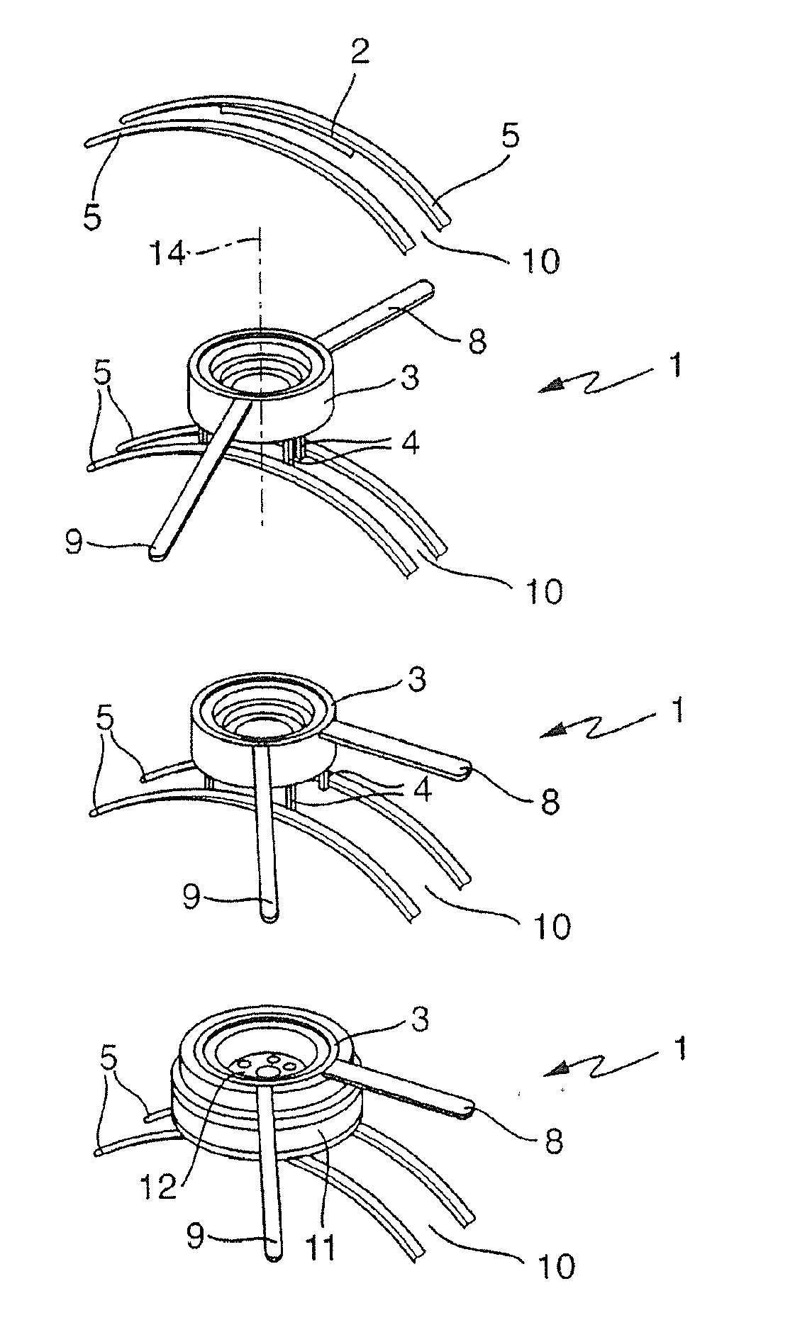
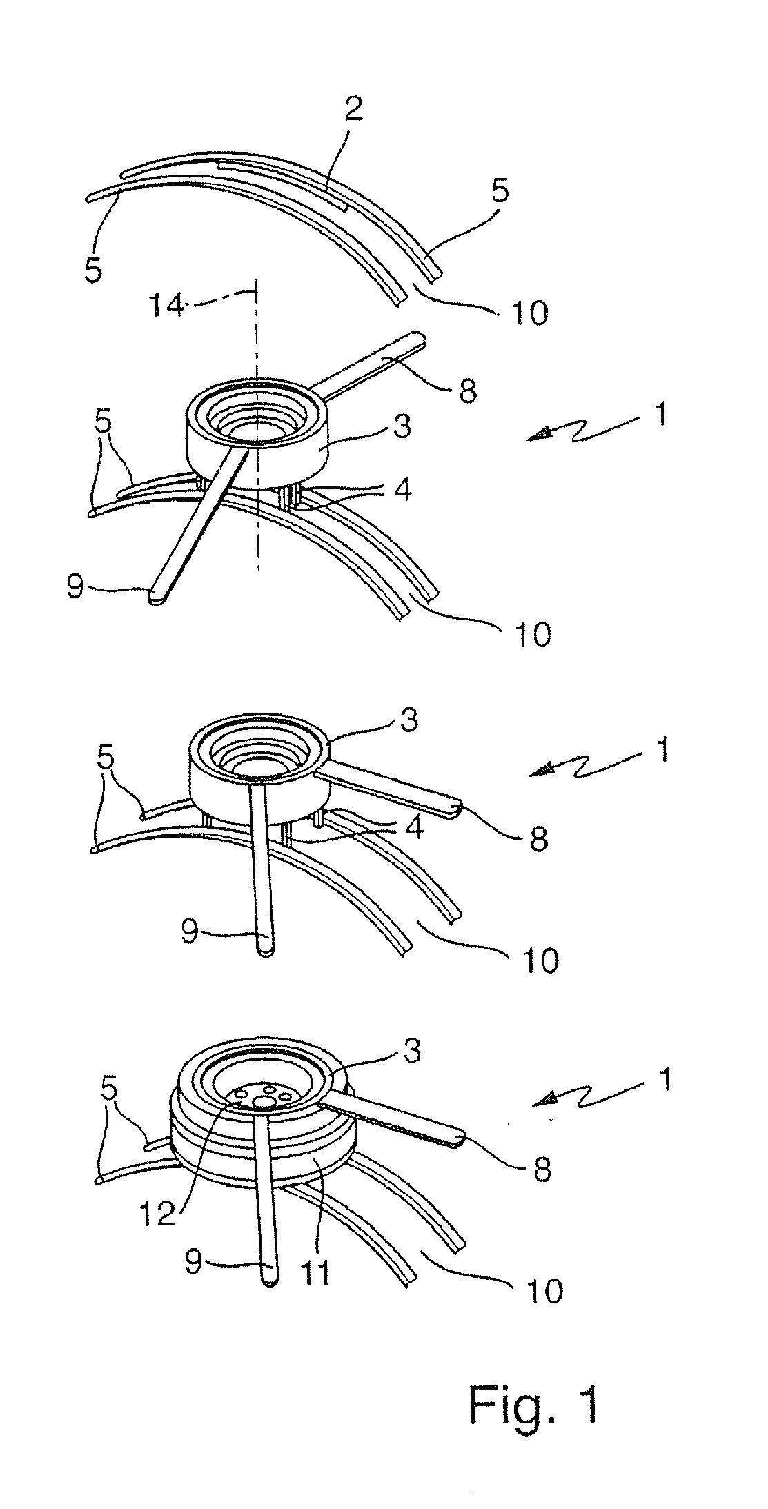
  • Device For Creating An Intercostal Transcutaneous Access To An, In Particular Endoscopic, Operating Field
  • Device For Creating An Intercostal Transcutaneous Access To An, In Particular Endoscopic, Operating Field
  • Device For Creating An Intercostal Transcutaneous Access To An, In Particular Endoscopic, Operating Field

Examples


first embodiment

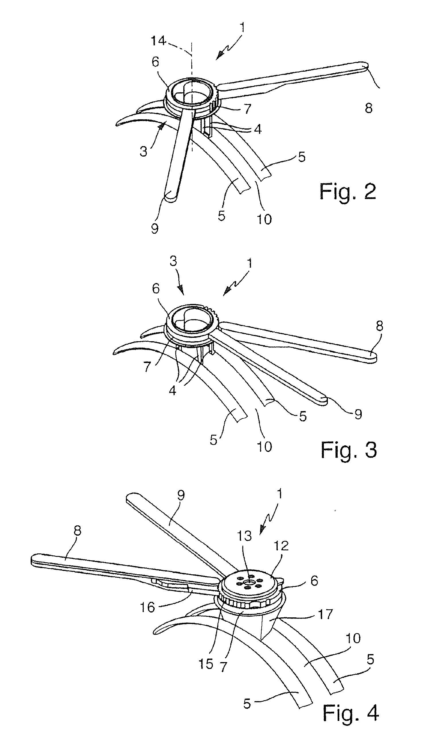
[0046]As can be seen from figures FIGS. 2 to 4, the base body 3 for this first embodiment of a device 1 for creating an intercostal transcutaneous access to an operating field comprises two housing sections 6 and 7 formed as concentric rings which can be turned relative to each other about a vertical axis 14 by means of the handles 8 and 9. When pressing together the handles 8 and 9 the housing sections 6 and 7 are turned against each other in such a way that the webs 4 mounted on the housing sections 6 and 7 are moved apart starting from their alignment in a straight line in the insertion position, as can be seen in the sequence of figures FIGS. 2 and 3.

[0047]Due to the arrangement of the webs 4 on the respective housing section 6 and 7 the webs 4 span in each, the intercostal space 10 spreading operating position of the webs 4 a parallelogram-shaped plane between each other, forming an access to the operating field.

[0048]The webs 4 can be locked relative to each other in their res...

third embodiment

[0062]The third embodiment shown in figures FIGS. 7 and 8 to create the device 1 essentially differs from both of the previously described embodiments in that the two housing sections 6 and 7 which form the base body 3 are pivotable about a horizontal axis 23 relative to each other.

[0063]For the embodiment shown, each housing section 6 and 7 of the base body 3 has only one web 4, which extends over the whole width of the base body 3. When pressing apart, the handles 8 and 9 shown in FIG. 8 the housing sections 6 and 7 are pivoted against each other about the horizontal axis 23 by means of which the webs 4 firmly connected to the housing sections 6 and 7 are pressed outwards thereby pressing the ribs 5 apart and increasing the intercostal space 10.

[0064]The devices 1 described above formed to create an intercostal transcutaneous access to an operating field are characterized in that the webs 4 are adjustable relative to each other which means that the size of intercostal space 10 can...


PUM


Abstract

A device for creating an intercostal transcutaneous access to an, in particular endoscopic, operating field including a base body, which closely surrounds an incision all-side frame-shaped and with at least two websmounted on the base body, which can be introduced into an intercostal space, by which two ribs arranged next to each other can be moved apart. To create a device for creating an intercostal transcutaneous access to an, in particular endoscopic, operating field which allows individual pressing apart of the ribs, it is proposed according to the invention that the at least two webs are mounted adjustable relative to each other on the base body.

Description

FIELD OF THE INVENTION[0001]The invention relates to a device for creating an intercostal transcutaneous access to an, in particular endoscopic, operating field comprising a base body, which closely surrounds an incision all-side frame-shaped and with at least two webs mounted on the base body, which can be introduced into an intercostal space, by which the two ribs arranged next to each other can be moved apart.BACKGROUND OF THE INVENTION[0002]During thoracic surgery it is necessary to gain access to the operating field intercostally, that is between the ribs of the patient. For open surgery a large incision is made and the operation is performed using clamps to pry the ribs apart, wherein it is not uncommon that the ribs break under the influence of such a spreading technique.[0003]An alternative to the classic open surgery technique is minimal invasive thoracic endoscopy (thoracocscopy), for which a multiplicity of small access points are created between the ribs through which th...

Claims


Application Information

Patent Timeline
IPC IPC(8): A61B17/34A61B17/02
CPCA61B17/3423A61B17/0218A61B17/0206A61B2017/3488A61B2017/3445A61B2017/3419A61B2017/3427A61B17/0293A61B2017/3484
Inventor CUSCHIERI, ALFREDCOLEMAN, STUARTMARTIN, DUNCAN
Owner UNIVERSITY OF DUNDEE
Learn More