Digital fluid sample separation apparatus and methods for one-step quantitative sample analysis

a fluid sample and digital technology, applied in biochemistry apparatus and processes, fluid controllers, laboratory glassware, etc., can solve the problems of high cost of testing, high cost of equipment, and high cost of electrical sources and bulky equipment, so as to reduce the cost of testing and simplify the assay

Active Publication Date: 2016-08-11
RGT UNIV OF CALIFORNIA
View PDF2 Cites 6 Cited by
  • Summary
  • Abstract
  • Description
  • Claims
  • Application Information

AI Technical Summary

Benefits of technology

The patent text discusses the development of new techniques for testing substances using enzyme-linked immunosorbent assay (ELISA) based methods. These techniques offer a cost-effective alternative to traditional testing methods but require significant manual handling time. The text also mentions the use of lateral flow strips which can detect NA but require multiple manual steps and provide only qualitative detection. The technical effect of the patent text is to provide a more efficient and cost-effective way to test substances using advanced techniques that offer both quantitative and qualitative detection.

Problems solved by technology

The patent text discusses the current methods used for quantitative nucleic acid detection in body fluid samples and the limitations of each. The technical problem addressed is the need for a faster, more cost-effective, and simpler method that can combine rapid sample preparation and quantitative assay endpoint readout in the same diagnostic chip.

Method used


Image

  • Digital fluid sample separation apparatus and methods for one-step quantitative sample analysis
  • Digital fluid sample separation apparatus and methods for one-step quantitative sample analysis
  • Digital fluid sample separation apparatus and methods for one-step quantitative sample analysis

Examples


example 1

[0068]As an example of blood based disease NA detection, HIV viral load quantification in blood was used as a demonstration. To demonstrate digital NA amplification, Recombinase Polymerase Amplification (RPA) was used, which is a ˜40° C. isothermal NA amplification technique. With 30 minutes of incubating at 40° C. using reusable instant heat packs, on-chip detection of HIV-1 RNA from spiked blood samples was achieved. The quantitative digital nucleic acid (NA) detection dynamic range was 103-106 copies / ml.

[0069]RPA was chosen because of the relatively low incubation temperature that is required. The lower incubation temperature greatly reduces the risk of generating air bubbles, in contrast to using PCR, which heats the samples up to 95° C. RPA is also the fastest isothermal amplification method commercially available to date. This, in combination with its robustness when used with plasma samples, made it an ideal NA amplification technique to integrate with the DS chip. It should ...

example 2

[0087]This assay was performed to demonstrate that digital separation can integrate sample preparation with digital isothermal amplification using RPA, to detect Methicillin-resistant Staphylococcus aureus (MRSA) DNA directly from human whole blood samples in 30 minutes.

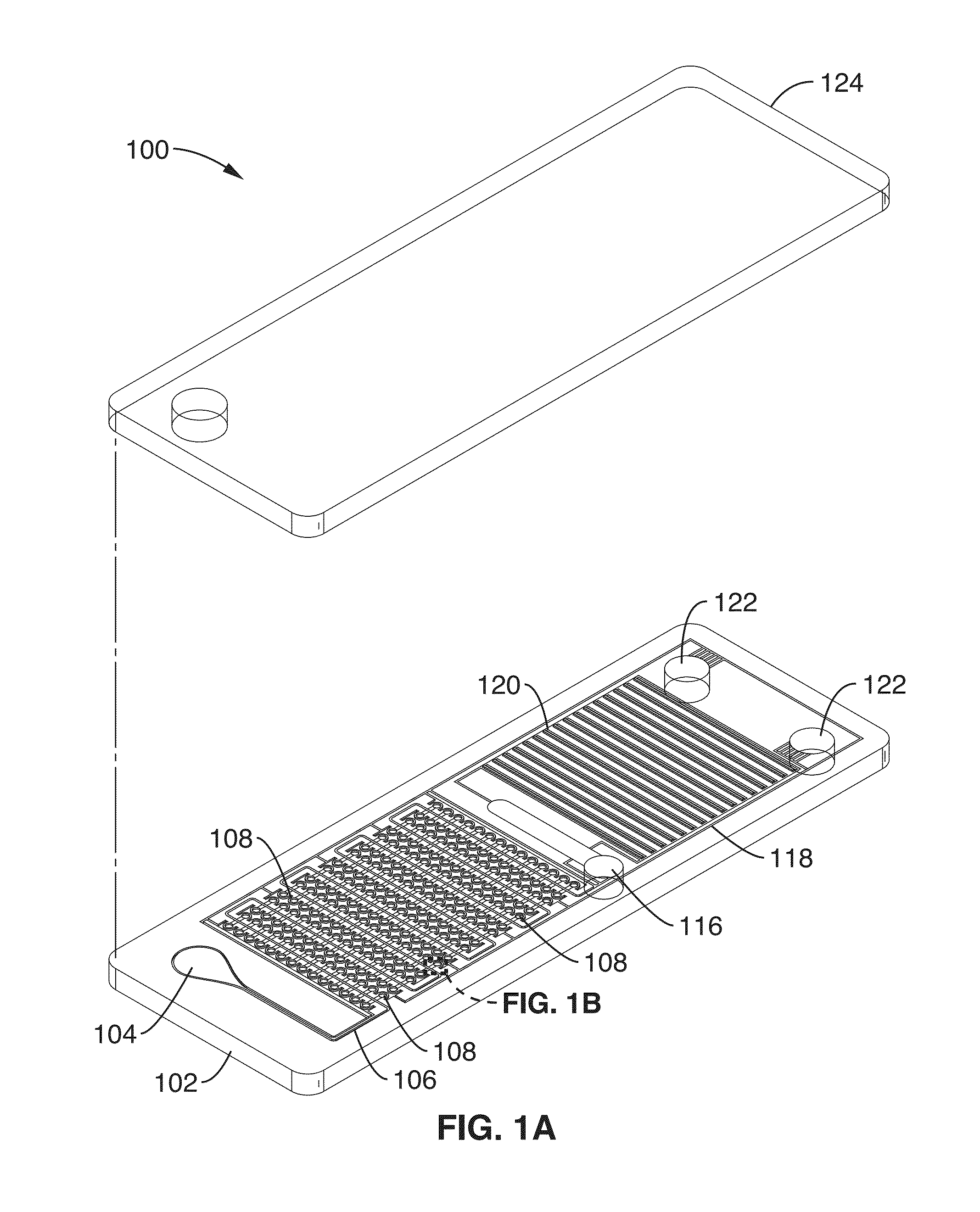
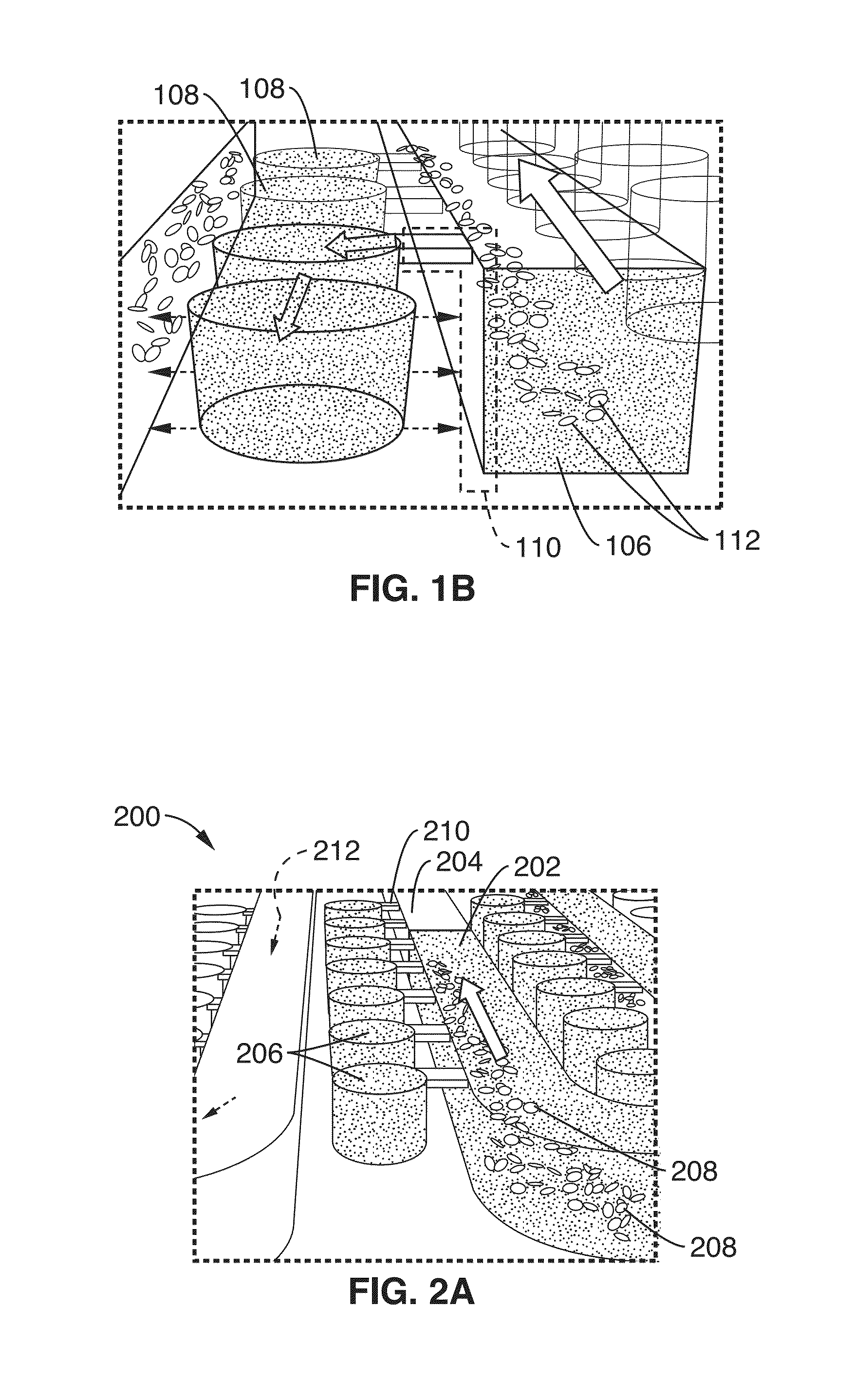
[0088]Using the cliff structures described in FIG. 1B, blood cell sedimentation was also used in this example to skim plasma into the wells. By separating the red blood cells, there is less optical obstruction of the fluorescence signal and less enzymatic interference of the polymerase due to hemoglobin inhibition. Since there is very low shear stress created on the red blood cells, there is minimal hemolysis. The DS chip design also avoids blood cells clogging since there are no features that would cause blood cell stacking against the flow. DS enables digital NA amplification assays (e.g. RPA) to be performed directly from separated plasma or other sample. In this example, the cliff structures and wells were arrang...


PUM

PropertyMeasurementUnit
Lengthaaaaaaaaaa
Lengthaaaaaaaaaa
Timeaaaaaaaaaa

Abstract

A Digital Separation (DS) chip for separating, digitizing and analyzing a fluid sample is presented. The DS chip includes a fluidic layer that prepares and compartmentalizes the fluid sample for analysis. Cliff structures that are adjacent to wells skim the fluid sample and prevent particles, which may interfere with fluid sample analysis, from entering the wells. Skimmed fluid sample analysis occurs in the wells and endpoint data can be collected and used to determine an original concentration of a desired component in the fluid sample very quickly. Using the described apparatus and methods, a fluid sample can be prepared, digitized, compartmentalized, assayed and the endpoint data collected in ˜30 minutes. The apparatus and methods can easily be adapted to provide parallel processing of a sample.

Description


Claims


Application Information

Patent Timeline
Owner RGT UNIV OF CALIFORNIA
Why Eureka
Try Eureka