
Cell image evaluation device, method, and program

a cell image and evaluation device technology, applied in image enhancement, instruments, microscopic object acquisition, etc., can solve the problems of inability to appropriately evaluate the state of the stem cell colony, and the possibility of appropriately evaluating

Active Publication Date: 2016-11-17
FUJIFILM CORP
View PDF16 Cites 7 Cited by
  • Summary
  • Abstract
  • Description
  • Claims
  • Application Information

AI Technical Summary

Benefits of technology

The present invention provides a cell image evaluation device, method, and non-transitory computer readable recording medium storing a program that can appropriately evaluate the state of a stem cell colony by taking into consideration the different changes in form of the respective local regions of the cells in the colony. This is achieved by acquiring specific information of a local region in the colony region of the cells in the cell image and determining an evaluation method for the cell image in the local region based on the specific information. The cell image evaluation device can determine an evaluation method for each local region based on the specific information and the information regarding the maturity of the cells. This allows for a more accurate evaluation of the stem cell colony.

Problems solved by technology

In the case of evaluating the state of a stem cell colony according to such locally different changes in the stem cell colony, for example, using the same evaluation method for the entire stem cell colony, the state of the stem cell colony may not be able to be appropriately evaluated.
In such a case, if the entire stem cell colony is evaluated using the form of individual cells, it is not possible to appropriately evaluate the stem cell colony since it is very difficult to recognize individual cells in the central portion due to lamination.

Method used


Image

  • Cell image evaluation device, method, and program
  • Cell image evaluation device, method, and program
  • Cell image evaluation device, method, and program

Examples


Embodiment Construction

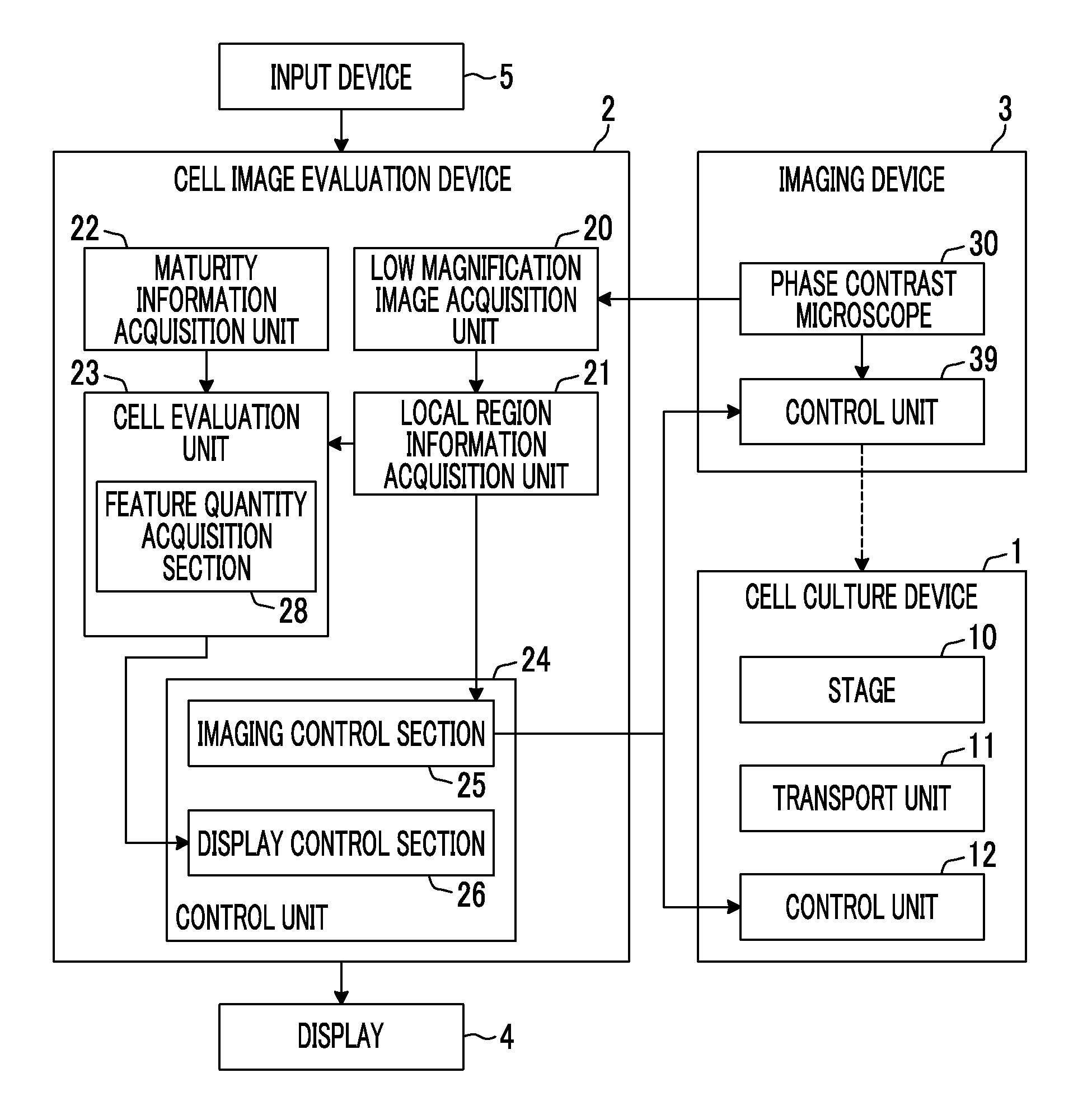
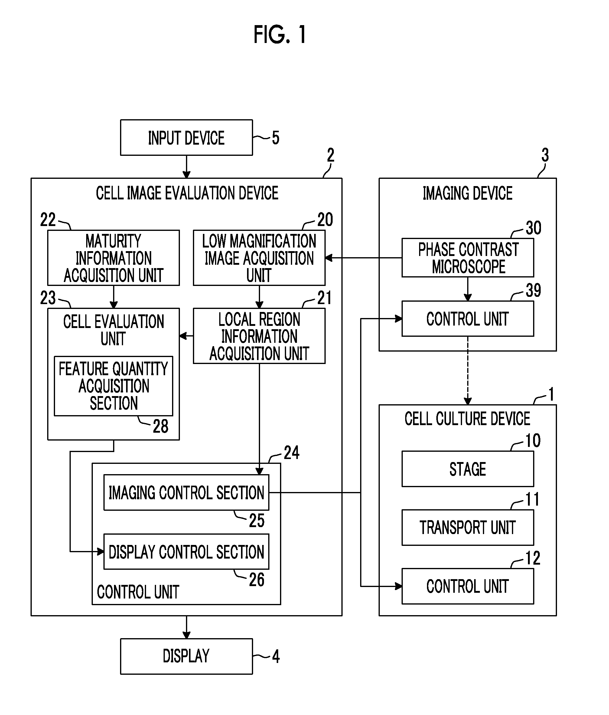
[0036]Hereinafter, an embodiment of a cell image evaluation device, method, and non-transitory computer readable recording medium storing a program of the present invention will be described in detail with reference to the diagrams. First, the overall configuration of a cell culture observation system including an embodiment of the cell image evaluation device of the present invention will be described. FIG. 1 is a block diagram showing the schematic configuration of a cell culture observation system.

[0037]As shown in FIG. 1, the cell culture observation system of the present embodiment includes a cell culture device 1, a cell image evaluation device 2, an imaging device 3, a display 4, and an input device 5.

[0038]The cell culture device 1 is a device for culturing cells. As cells to be cultured, for example, there are pluripotent stem cells such as iPS cells, ES cells, or STAP cells, cells of nerves, skin, myocardium, or liver that are differentiation-induced from stem cells, and c...


PUM


Abstract

There is provided a cell image evaluation device, method, and program to appropriately evaluate the state of a stem cell colony according to different changes in form of respective local regions of the cell colony. There are included a low magnification image acquisition unit 20 that acquires a cell image by imaging cells; a cell evaluation unit 23 that evaluates the cell image; and a local region information acquisition unit 21 that acquires the specific information of a local region in a colony region of the cells in the cell image. The cell evaluation unit 23 determines, for each local region in the colony region, an evaluation method for a cell image in the local region based on the specific information of the local region, and evaluates the cell image of the local region using the determined evaluation method.

Description

CROSS-REFERENCE TO RELATED APPLICATIONS[0001]This application is a Continuation of PCT International Application No. PCT / JP2015 / 051394 filed on Jan. 20, 2015, which claims priority under 35 U.S.C §119(a) to Patent Application No. 2014-043254 filed in Japan on Mar. 5, 2014, all of which are hereby expressly incorporated by reference into the present application.BACKGROUND OF THE INVENTION[0002]1. Field of the Invention[0003]The present invention relates to a cell image evaluation device, method, and non-transitory computer readable recording medium storing a program for evaluating cell image obtained by imaging cells.[0004]2. Description of the Related Art[0005]Conventionally, a method of imaging pluripotent stem cells such as ES cells, iPS cells, and STAP cells, differentiation-induced cells, or the like using a microscope or the like and determining the culture state of cells by ascertaining the features of the image has been proposed.[0006]For example, JP2011-239778A and JP2011-22...

Claims


Application Information

Patent Timeline
Patent Type & Authority Applications(United States)
IPC IPC(8): G06T7/00G06K9/00
CPCG06T7/0012G06K9/00134G06T7/0085G06T2207/10056G06K9/0014G06T2207/30024G06T7/0051G06T7/62G06V20/693G06V20/698G06V20/695
Inventor MATSUMOTO, TSUYOSHI
Owner FUJIFILM CORP
Learn More