Knowledge-based ultrasound image enhancement

Active Publication Date: 2016-12-01
SIEMENS MEDICAL SOLUTIONS USA INC
View PDF14 Cites 43 Cited by
  • Summary
  • Abstract
  • Description
  • Claims
  • Application Information

AI Technical Summary

Benefits of technology

[0006]In a second aspect, a non-transitory computer readable storage medium has stored therein data representing instructions executable by a programmed processor for image enhancement in medical diagnostic ultrasound. The storage medium includes instructions for receiving, from an ultrasound scanner, detected ultrasound data representing a patient, classifying locations represented by th

Problems solved by technology

Traditional image enhancement algorithms are limited in their ability to perfect an ultrasound image.
In many cases, imaging artifacts have the same or similar properties to anatomic structures or tissue and hence are not detectable and effectively segmenta

Method used


Image

  • Knowledge-based ultrasound image enhancement
  • Knowledge-based ultrasound image enhancement
  • Knowledge-based ultrasound image enhancement

Examples


Example

DETAILED DESCRIPTION OF THE DRAWINGS AND PRESENTLY PREFERRED EMBODIMENTS

[0016]Knowledge-based enhancement of ultrasound images is provided. Knowledge-based feature detection techniques may successfully detect anatomic structures or artifacts in an ultrasound image without detecting other objects. These detection techniques are harnessed to improve and make smarter image processing or enhancement algorithms. Post-acquisition image enhancement benefits from application of knowledge-based detection. Already acquired image data may be altered by image processing localized specifically to detected anatomy or artifacts.

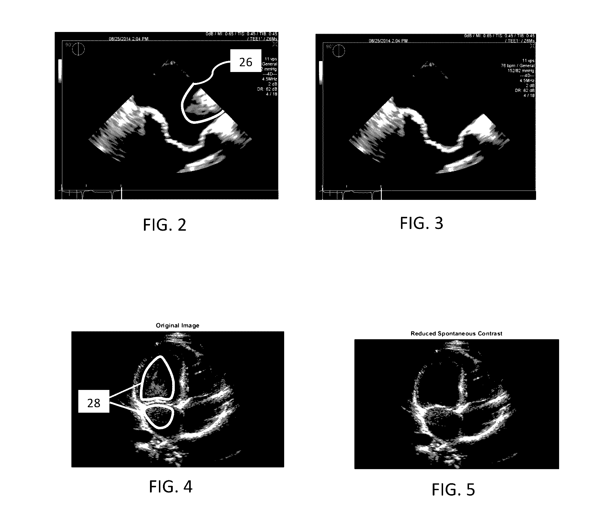
[0017]In one embodiment, imaging artifacts are identified for enhanced imaging. The knowledge is captured as expert user annotations of artifacts in a knowledge database. This knowledge significantly improves artifact detection and minimization. Utilizing knowledge-based detection algorithms to detect artifacts, such as grating lobes, side lobes from rib reflections, rib sh...


PUM


Abstract

Image enhancement is provided for medical diagnostic ultrasound. Knowledge-based detection of anatomy or artifact identifies locations to be enhanced. The knowledge-based detection of the locations may avoid identification of other anatomy or artifacts. The image enhancement is applied to the identified locations and not others.

Description

BACKGROUND[0001]The present embodiments relate to medical diagnostic ultrasound imaging. In particular, ultrasound imaging is enhanced.[0002]Traditional image enhancement algorithms are limited in their ability to perfect an ultrasound image. The image enhancement operates without specific prior knowledge of the anatomy and pathology represented in the image. Basic image processing techniques, such as low pass filters, reduce speckle noise but blur out anatomic structures.[0003]In many cases, imaging artifacts have the same or similar properties to anatomic structures or tissue and hence are not detectable and effectively segmentable by image processing algorithms. The image processing may even enhance the artifacts. High pass filters used for edge enhancement enhance speckle noise. Techniques that are more complex rely on standard image analysis, such as gradient, variance, or simply amplitude-based image segmentation to process selectively various parts of the image. While these t...

Claims


Application Information

Patent Timeline
IPC IPC(8): G06K9/62A61B8/13G06K9/46G06T7/00G06T5/00A61B8/00A61B8/08G06V10/774
CPCG06K9/6256A61B8/463A61B8/13A61B8/0891G06T7/0012G06T2207/10132G06K9/6267G06K9/4661G06T7/004G06T2207/30101G06T5/00A61B8/52A61B8/5269G06T5/006G06T2207/20081G06T2207/20004G06T2207/20012G06T2207/20076G06T2207/20192G06T2207/30004G06V10/774A61B8/5207A61B8/5215G06T5/001G06F18/214
Inventor RAO, BIMBAHOULE, HELENEGEORGESCU, BOGDAN
Owner SIEMENS MEDICAL SOLUTIONS USA INC
Why Eureka
Try Eureka