Though fine textural changes in tissue have long been recognized as the earliest markers in a wide range of diseases, robust clinical assessment of fine texture remains elusive, the main difficulty arising from blurring caused by subject motion over the time required for
data acquisition.
Changes in fine tissue texture have been recognized for many years by diagnosticians, including radiologists and pathologists as the earliest harbinger of a
large range of diseases, but
in vivo assessment and measurement of fine texture has remained outside the capabilities of current imaging technologies.
But post
processing analysis is limited in effect as it doesn't deal with the underlying problem that prevents
high resolution acquisition of textural information, i.e. subject motion.
However, even with gating, there is sufficient variation between acquisitions to cause loss of spatial
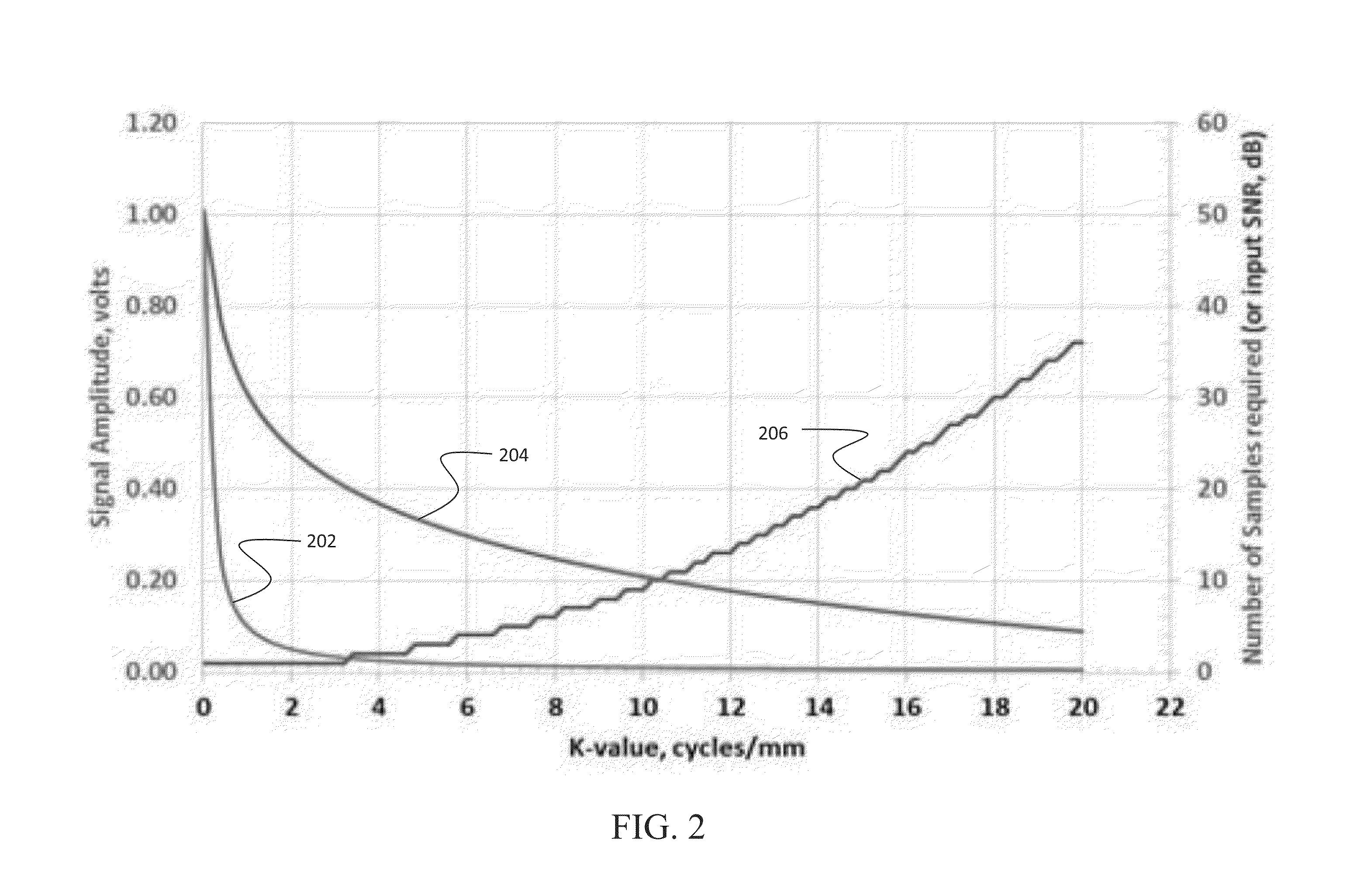
phase coherence at the high k-values of interest for texture measurements.
This problem is exacerbated by the fact that motion may not be perfectly cyclic, and often originates from combined sources.
Twitching is rapid, inducing random displacements, and hence it is not possible to maintain coherence at the high k-values of interest when measuring texture.
While
Positron Emission
Tomography (PET) provides valuable
diagnostic information, it is not capable of resolution below about 5 mm and relies on the use of radioactive tracers for imaging as well as x-
ray beams for positioning, raising
dose concerns, especially if repeat scanning is needed.
The non-negligible risk from the associated
radiation dose makes CT problematic for longitudinal imaging and limits available resolution.
Along with serious
dose concerns, digital x-
ray resolution is limited because the 2-dimensional image obtained is a composite of the absorption through the entire thickness of tissue presented to the beam.
Current clinical diagnostics for the diseases that are the target of the method claimed herein are fraught with difficulties in obtaining sufficient
in vivo resolution, or accuracy.
In some cases, no definitive diagnostic exists currently.
In other pathologies, particularly in breast and liver, diagnosis is dependent on
biopsy, with its non-negligible risk of morbidity and even mortality, and which is prone to high read and sampling errors.
But, though TBS does not yield a detailed assessment of trabecular architecture.
In MRI, though
high contrast between bone and marrow is readily obtained, resolution is limited by
patient motion over the long time needed to acquire an image with sufficient resolution to characterize the trabecular network.
Further, a large
data matrix, hence long
acquisition time, is still required to obtain sufficient image information to determine trabecular spacing and element thickness.
This long
acquisition time results in varying levels of motion-induced blurring, depending on
patient compliance-twitching is still a serious problem even when measuring extremities.
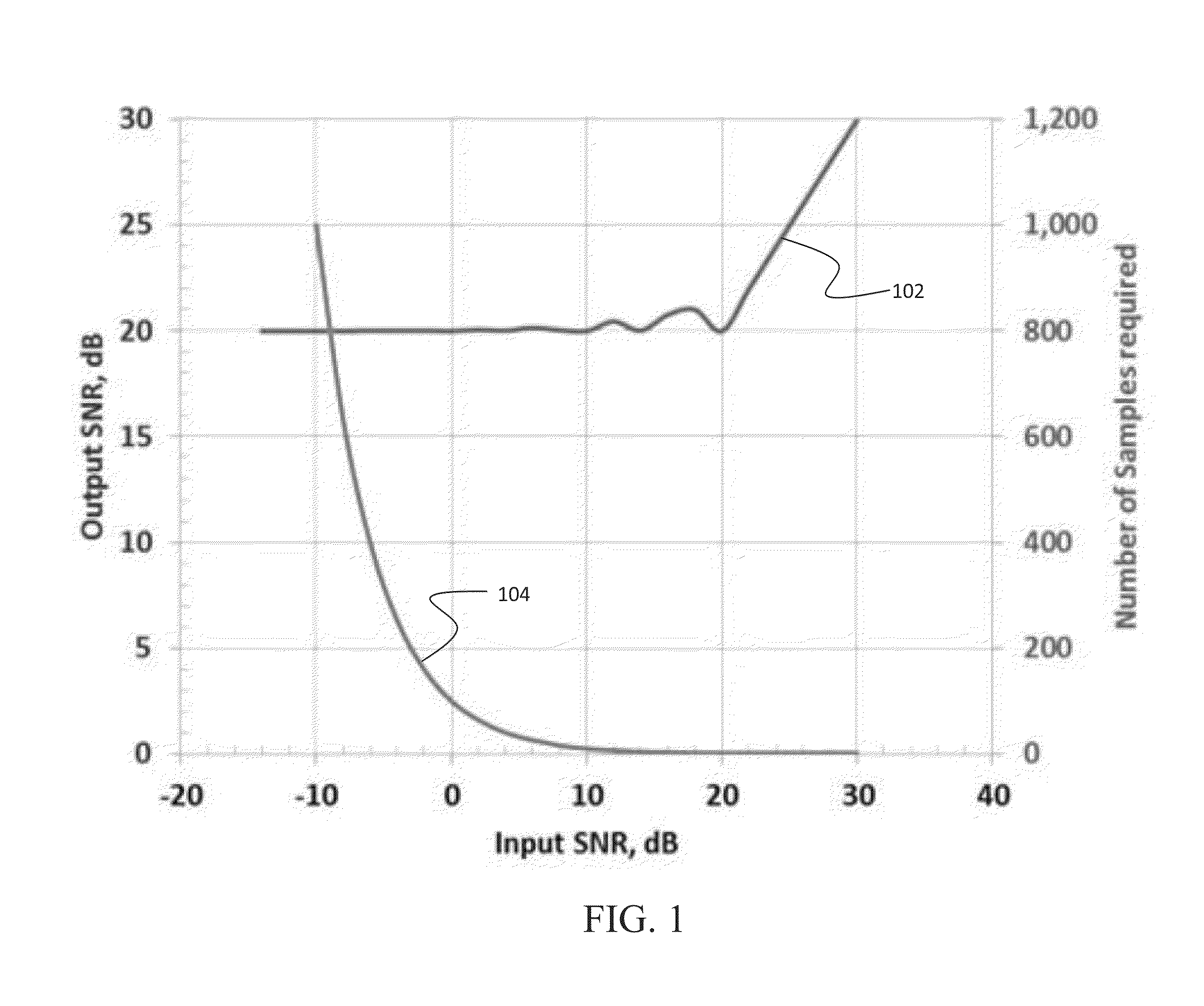
However, as the gradient encoded echoes, are very low
Signal to
Noise (SNR),
noise averaging is required.
Until now, no clinical technique has been able to provide this capability.
Currently, the
gold standard for
pathology assessment is
tissue biopsy—a highly invasive and often painful procedure with a non-negligible morbidity—and mortality—risk (patients need to stay at the hospital for post-
biopsy observation for hours to overnight), and one that is prone to sampling errors and large reading variation.
Magnetic
Resonance-based
Elastography (MRE), which has been under development for some time for use in assessment of
liver disease, is not capable of early-stage assessment—the read errors are too large prior to significant fibrotic invasion (advanced disease).
Further, this technique requires expensive additional hardware, the presence of a skilled
technician, and takes as much as 20 minutes total set up and scanning time, making it a very costly procedure.
The ability to image fibrotic texture directly by
MR imaging is compromised both by
patient motion over the time necessary to acquire data and by lack of contrast between the fibers and the surrounding tissue.
Even acquisition during a
single breath hold is severely compromised by cardiac pulsatile motion and noncompliance to breath hold, which results in significant motion at many organs, such as liver and lungs.
And SNR is low enough that
motion correction by combining reregistered MR-intensity profiles obtained from successive echoes is extremely problematic.
Similarly, assessment of the amount of
cardiac fibrosis in early stage disease using MRI is seriously hampered by cardiac pulsation over the time of the measurement.
However, ability to assess such changes in the brain is only available post mortem.
Various
in vivo diagnostic techniques are available for AD and other dementias, but none of them are definitive.
As discussed previously,
PET imaging is extremely expensive, cannot provide
high resolution, and relies on use of radioisotopes and positioning x-
ray beams, complicating approval for longitudinal use due to dose concerns.
Use of CSF biomarkers for
dementia diagnosis is painful and highly invasive and cannot differentiate
signal levels by anatomic position in the brain, as is possible with imaging biomarkers.
As various forms of
dementia are found to have different spatial / temporal progression through the brain, this is a serious drawback to use of
liquid biopsy.
This textural size, and the fact that the cortex is extremely thin, makes speed of acquisition paramount, as even tiny patient motion will make data collection impossible.
However, in order to circumvent the problem of
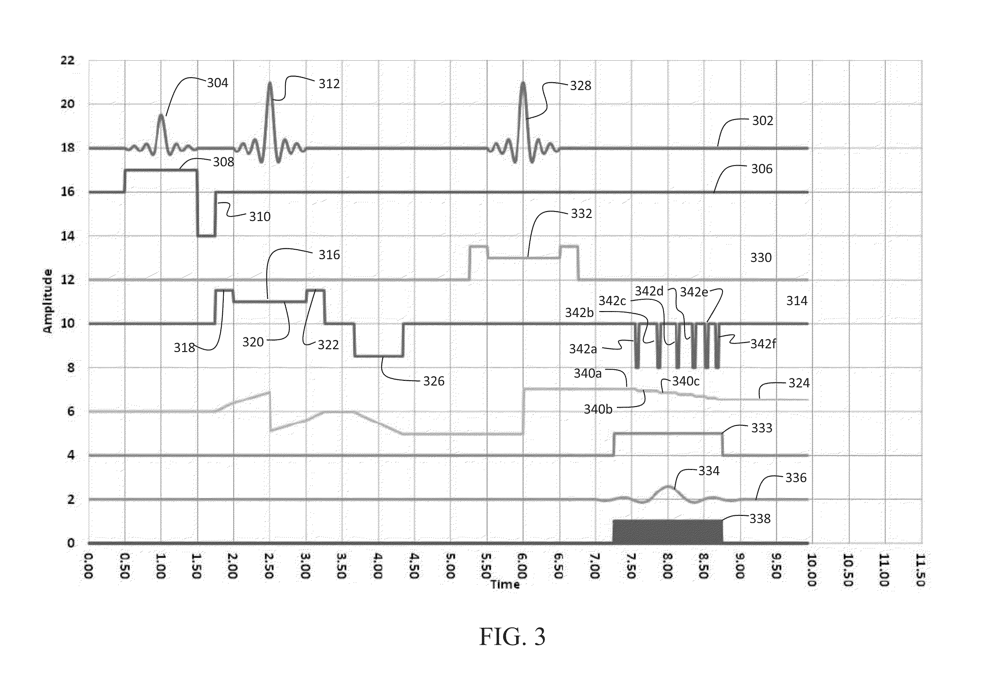
signal degradation due to patient motion, data must be taken on a time scale not previously possible.


