
Prevention of Ventilator Associated Pneumonia (VAP)

a technology of ventilator and associated pneumonia, which is applied in the field of patient treatment, can solve the problems of increased morbidity and mortality, difficult and expensive diagnosis of vap, and the cost of diagnosing and treating vap exceeds 1.1 billion dollars annually, and achieves the effect of preventing vap

Active Publication Date: 2017-01-12
VIRGINIA COMMONWEALTH UNIV
View PDF9 Cites 0 Cited by
  • Summary
  • Abstract
  • Description
  • Claims
  • Application Information

AI Technical Summary

Benefits of technology

The anti-VAP system effectively reduces the incidence of VAP by controlling the space where secretions accumulate, providing a physical or absorptive barrier to prevent microaspiration and colonization, thereby decreasing the risk of pneumonia and associated complications.

Problems solved by technology

VAP may occur in up to 65% of patients in the intensive care unit (ICU) and is associated with an increase in morbidity and mortality.
It is estimated that cost of diagnosing and treating VAP exceeds 1.1 billion dollars annually.
The diagnosis of VAP is difficult and expensive.
Controversy continues to exist in the methodology in making a definitive diagnosis.
Treatment is also controversial and the use of empiric antibiotics is believed to have contributed to making the overall treatment of true VAP more difficult.
A major cost to the treatment is prolonging the time patients require mechanical ventilation and thus care in the ICU setting.
This increased time of treatment in this setting is likely to actually increase the chances of additional complications including developing additional VAPs and antibiotic resistant organisms.
However, higher volumes will result in higher pressures being created between the cuff and the tracheal mucosa thus placing the tracheal mucosa at risk for necrosis.
The disadvantage of this method is that suction is required.
Because endotracheal intubation occurs in many non-ICU areas, suction is not readily available.
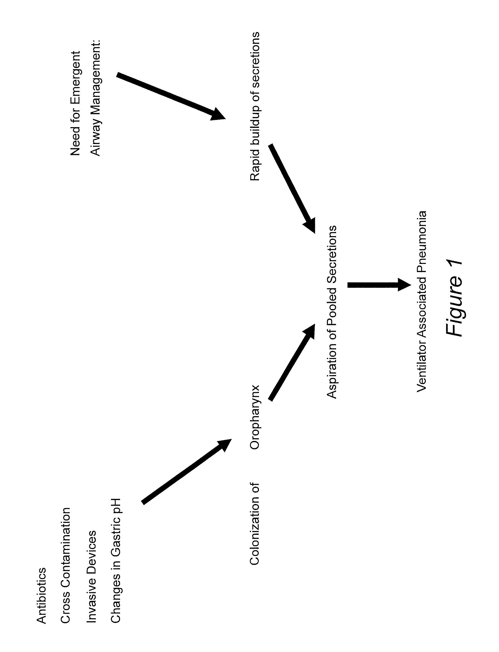
Patients are likely to be a most risk for aspiration from subglottic secretions very early after intubation especially when it is performed in less than ideal places such as on the wards, the emergency department, or in the prehospital setting.
A number of methods, which involve changes in cuff design, have reported various degrees of success but none have undergone extensive clinical testing.
In regards to this, intubation done in less than ideal circumstances where patients may be at greatest risk for microaspiration means that antibiotic embedded systems or tubes designed to prevent formation of biofilm might not be effective in this time early time range.
Another major problem in the patient requiring endotracheal intubation and mechanical ventilation is the need for sedation due to the coughing reflexes induced by contact of the endotracheal tube and cuff with points of the supra and subglottic portions of the larynx.
This degree of additional sedation can impede physical and neurologic assessment of the patient and delay efforts for weaning of mechanical ventilation.
This is of great importance because additional time spent utilizing mechanical ventilation will necessarily result in the incurrence of significant expense and may potentially result in the development of VAP with all of its complications and additional expense.
Conventional strategies to reduce VAP necessitate purchase of a separate endotracheal tube, which making implementation difficult especially if VAP prevention strategies are to be performed in all settings at the earliest possible time.
In addition, each conventional strategy is relatively singular or limited in its ability to prevent VAP.
However, this manual hygiene work on intubated patients is labor-intensive, and even so some of them still develop VAP.
Use of such techniques in the early stages of intubation such as in the pre-hospital and emergency department settings is not practical.
This is unfortunate because these are places where patients are at major risks and where the normal microbial oral flora of the patient rapidly changes to more virulent hospital-based pathogens.

Method used


Image

  • Prevention of Ventilator Associated Pneumonia (VAP)
  • Prevention of Ventilator Associated Pneumonia (VAP)
  • Prevention of Ventilator Associated Pneumonia (VAP)

Examples


example 1

Leakage Experiment

[0062]Experimentation regarding anti-VAP devices and / or anti-VAP materials was performed as follows.

[0063]7 mm ETT inflated in the barrel of 20cc syringe, with dye leaking occurring around the cuff through channels formed between the cuff and the balloon: A photograph was taken that shows a screening methodology reported in other studies that uses the barrel of a 20 cc syringe to act as the trachea. It is intubated with an endotracheal tube followed by inflation of the cuff and introduction of dye above the balloon. A 7 mm ETT was used. Leakage of dye around the balloon can then be observed for. Leakage of dye is seen when the balloon is filled with 10 cc air.

[0064]7 mm ETT with rubber latex cover around the balloon, according to an embodiment of the invention; native balloon inflated: In another photograph, another 7 mm ETT is used but before insertion into the “trachea” it was placed through a simple piece of a latex rubber drain. Inflation of the native balloon ...

example 2

[0065]FIG. 3 shows an inventive open ended sleeve 500 or condom to be placed over an ETT prior to intubation. A portion 501 of the sleeve 500 goes over the ETT balloon and expands as the balloon expands.

[0066]Modification of the end of the sleeve 500 allows for the native balloon to be covered with a material that would not lead to formation of channels between the ETT attachment and the tracheal mucosa when the native balloon is inflated. The sleeve 500 may be embedded with antimicrobials / bacteriostatic agents and anaesthetics. Materials used for forming the sleeve 500 preferably are resistant to formation of biofilms.

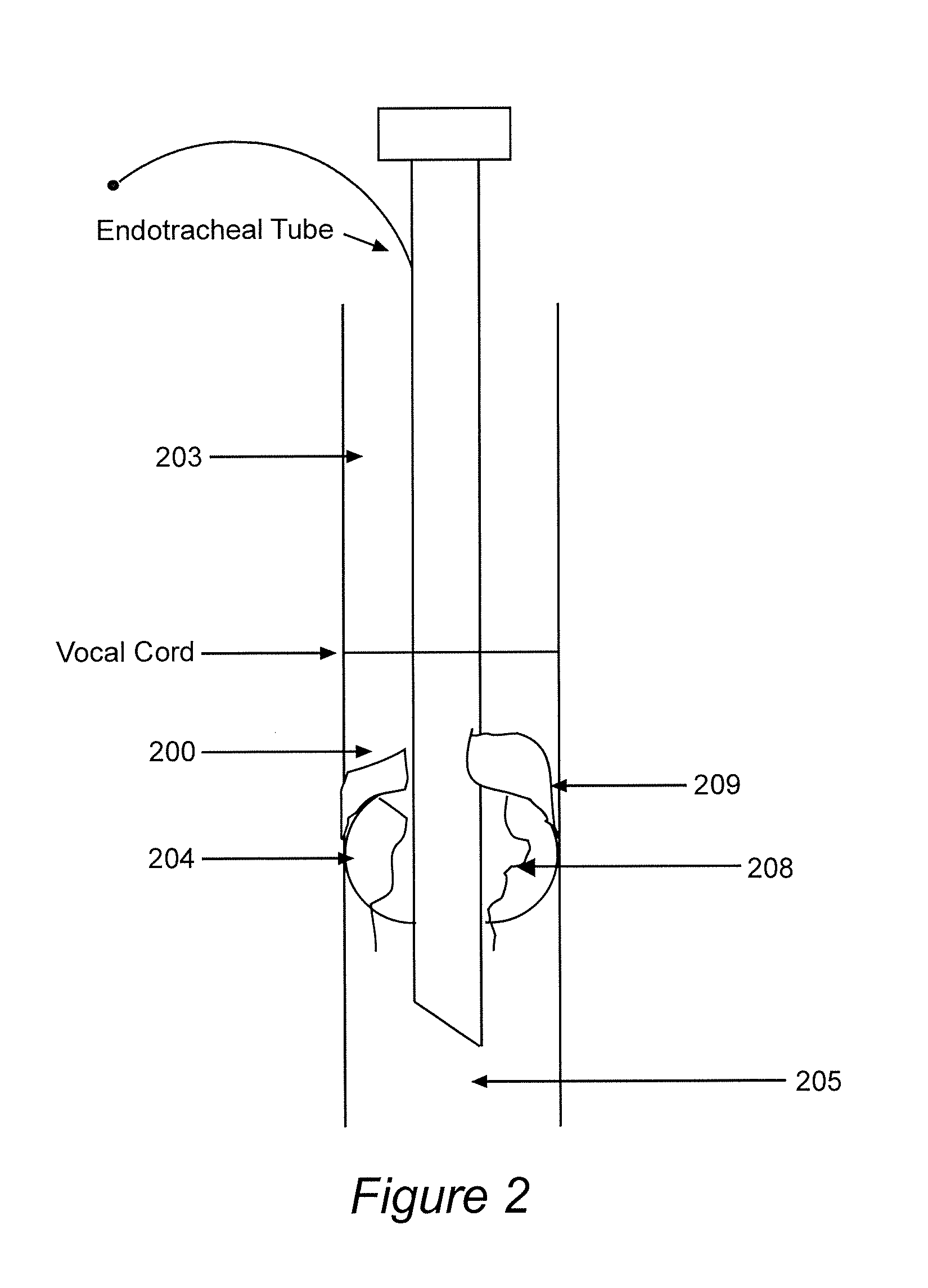
[0067]FIG. 4 shows the inventive sleeve 500 extending from below the balloon to the proximal tube when the sleeve 500 is used with a traditional endotracheal tube ETT with a pilot balloon for cuff inflation. The endotracheal tube ETT, supraglottic space 203, vocal cords VC, and subglottic space 200 in FIG. 4 are as in FIG. 2. The sleeve 500 optionally may have a separ...

example 2a

[0068]FIG. 5 is a modified version of the sleeve 500 of FIG. 3, modified to include ports 709 for delivery of compounds (such as antimicrobials, anesthetics, etc.) and port 708 for injection of compounds (such as anesthetics, antimicrobials, etc.).


PUM


Abstract

Ventilator associated pneumonia (VAP) may be prevented in a patient, or its occurrence reduced in a population of patients, by using an anti-VAP device or an anti-VAP material such as an anti-VAP mouthpiece that absorbs secretions. By reducing the problem of bacterial-containing secretions that otherwise build up in the airway of, and elsewhere in, the intubated patient, VAP can be prevented from occurring in intubated patients, such as patients intubated with an endotracheal tube (ETT) or a nasogastric tube. Anti-VAP mouthpieces also are useable in non-intubated patients to maintain oral hygiene.

Description

RELATED APPLICATIONS[0001]This is a continuation of U.S. application Ser. No. 13 / 181,843, filed Jul. 13, 2011, entitled “Prevention of Ventilator Associated Pneumonia (VAP)”, which is a continuation-in-part of U.S. application Ser. No. 11 / 217,667, filed Sep. 2, 2005, entitled “Prevention of Ventilator Associated Pneumonia (VAP)”, which claims the benefit of U.S. Provisional Application No. 60 / 607,070, filed Sep. 3, 2004, entitled “Device and Methods to Prevent Ventilator Associated Pneumonia and to Provide Laryngeal Anesthesia During Mechanical Ventilation.”FIELD OF THE INVENTION[0002]This invention relates to treatment of patients who are intubated with an endotracheal tube (ETT), and especially to ventilator associated pneumonia (VAP) undesirably associated with such intubation.BACKGROUND OF THE INVENTION[0003]Ventilator associated pneumonia (VAP) is a potentially preventable cause of pneumonia that occurs in patients who are endotracheally intubated and mechanically ventilated fo...

Claims


Application Information

Patent Timeline
Patent Type & Authority Applications(United States)
IPC IPC(8): A61M16/04
CPCA61M16/049A61M2205/0205A61M16/0434A61M16/0436A61M16/0479A61M16/04
Inventor WARD, KEVIN R.SESSLER, CURTIS N.GRAP, MARY JODINARDO, LAWRENCE J.SPIESS, BRUCE D.IVATURY, RAO R.MUNRO, CINDY
Owner VIRGINIA COMMONWEALTH UNIV
Learn More